移植肾功能减退患者肾组织中环孢素A测定及其意义
作者:凡杰 王祥慧 徐达 王跃闽 肖家全 刘永 倪灿荣 唐孝达
单位:凡杰(上海市第一人民医院泌尿科 上海,200080);王祥慧(上海市第一人民医院泌尿科 上海,200080);徐达(上海市第一人民医院泌尿科 上海,200080);王跃闽(上海市第一人民医院泌尿科 上海,200080);肖家全(上海市第一人民医院泌尿科 上海,200080);刘永(上海市第一人民医院泌尿科 上海,200080)
关键词:肾移植;免疫组织化学;环孢素
肾脏病与透析肾移植杂志000106 摘 要 目的:探讨免疫组织化学EnVision方法在移植肾功能减退患者肾组织中检测环孢素A(CsA)的可能性及其意义。 方法:收集我院1988年至1998年间肾移植术后长期服用CsA、血肌酐增高,临床表现为移植肾功能减退的患者11例,在B超引导下从移植肾上极、下极穿刺活检,正常肾组织作为对照组。采用免疫组织化学染色技术检测CsA及多药耐药P-gp蛋白在移植肾组织中的表达,观察其在组织切片中的形态学变化。 结果:本组11例肾移植患者中,有8例(72.7%)CsA阳性,对照组CsA为阴性;CsA以肾小管阳性率最高(72.7%),小血管次之(45.5%),肾小球最低(27.3%)。P-gp在移植肾组织中也以肾小管表达最高(81.8%),小血管次之(27.2%),肾小球最低(18.1%)。CsA和P-gp在移植肾组织中表达具有一致性,二者同时阳性以肾小管最高,共7例(63.6%)。患者血中环孢素浓度超过正常值的次数,与环孢素在移植肾组织中的阳性率不一致。肾移植后时间的长短与移植肾组织中CsA和P-gp表达程度不相关。 结论:检测移植肾肾组织中CsA的表达,有可能成为肾移植术后监测CsA肾毒性的一个重要指标,而同时检测多药耐药P-gp蛋白在移植肾组织中的表达,则可以提高诊断准确性。
, http://www.100md.com
CLINICAL SIGNIFICANCE OF CYCLOSPORINE A DETECTION IN RENAL ALLOGRAFTS
FAN Jie WANG Xianghui XU Da WANG Yaomin XIAO Jiaquan LIU Yong NI Canrong TANG Xiaoda
(Department of Urology,Shanghai First Peoples Hospital,Shanghai,200080)
OBJECTIVE To study the relationship between cyclosporine A nephrotoxicity and renal allograft dysfunction by detecting cyclosporine A(CsA)in renal allografts. METHODOLOGY Eleven patients on triple immunosuppressive therapy(CsA,axathioprien and steroid)with renal graft dysfunction after transplantation(increased serum creatinine>190 μmol/L)underwent allograft biopsy.Under the guidance of B mode ultrasound,a protocol biopsy of the renal graft was performed and autopsy tissue sections from normal kidney served as control group.The expressions of cyclosporine A and P-gp in renal allografts were detected by using immunohistochemical staining technique in 9 cases of chronic rejection,1 case of early acute rejection,and 1 case having no lesion in histological examination.Light microscopy examination and localization of CsA and P-gh in the graft specimen were evaluated with respect to the patterns of histologic lesion. RESULTS The expression of CsA was positive in eight out of 11 renal transplant recipients with allograft dysfunction,and was all negative in the control group.The incidence of the expression of CsA was 72.7%(8/11)in renal tubule,45.5%(5/11)in arteriol and 27.3%(3/11)in glomeruli respectively.The expression of P-gp in renal tubule was 81.8%(9/11),in arteriol 27.2%(3/11)and in glomeruli 18.1%(2/11)respectively.The incidence of the expression of CsA and P-gp(predominantly in renal tubule)was 63.6%(7/11).7 of 11 renal transplant recipients who underwent the protocol biopsy had CsA nephrotoxicity proved by histological examination. CONCLUSION Combined detection of cyclosporine A and P-gp is of clinical significance in the diagnosis of cyclosporine A nephrotoxicity.
, 百拇医药
Key words kidney transplantation immunohistochemistry cyclosporine A
环孢素A(CsA)肾毒性是急、慢性移植肾功能减退的原因之一,但CsA肾毒性常难以与其它影响移植肾功能减退的危险因素相鉴别。我们利用免疫组织化学的方法研究CsA在肾功能减退患者移植肾组织中的分布,以探索利用免疫组织化学方法在移植肾组织中检测CsA的可能性,为诊断CsA肾毒性提供新的方法。另外,CsA能够诱导肾脏等组织细胞多药耐药蛋白(P-gp)的表达增加[1],因此联合检测移植肾组织中CsA、P-gp的表达,则有可能提高诊断CsA肾毒性的准确性。
1 对象和方法
1.1 临床资料 我院1988年至1998年间肾移植患者11例,临床表现为移植肾功能减退,其临床资料见表1,所有病例均在我院接受长期随访。以4例肾癌手术切除后的肾组织无癌区,(病理证实为正常肾组织),作为对照组。对照组活检取材大小为0.4cm×0.6cm×0.7cm。实验组分别在B超引导下从移植肾上极、下极穿刺活检,组织块大小约为1.50cm×0.05cm×0.01cm,每例取2~3个组织块。活检组织用100 g/L的甲醛溶液固定,脱水,石蜡包埋,同时作HE染色。
, http://www.100md.com
1.2 试剂
1.2.1 P-gp混合单抗 JSB,C219,C494为丹麦DAKO公司产品,1∶50稀释。
1.2.2 环孢素A单抗 瑞士Norvatis公司产品。
1.2.3 其它试剂 EnVision Kit为丹麦DAKO公司产品。
1.3 实验方法 石腊切片常规脱腊,PBS缓冲液冲洗,微波抗原修复,然后用3%H2O2阻断内源性过氧化酶,PBS洗3×3min,依次加入一抗(CsA单抗或P-gp混合单抗),4℃过夜,PBS洗3×3min,再滴加EnVision多聚物酶复合物孵育37℃ 30min。0.05%DAB+0.03%H2O2显色,苏木素复染,逐级脱水,透明封片,光镜观察。用PBS代替一抗作阴性对照,HE染色明确病理诊断。
, 百拇医药
1.4 免疫组织化学染色结果判定
1.4.1 P-gp表达 ①:50%以上的肾小管、肾小球细胞及小血管呈阳性表达;②:25%~50%的肾小管、肾小球细胞及小血管呈阳性表达;③+:25%以下的肾小管、肾小球细胞及小血管呈阳性表达;④正常(-):内散在个别阳性细胞,一般少于10%。
1.4.2 CsA表达 ①:50%以上的肾小管、肾小球细胞及小血管阳性;②:25%~50%的肾小管、肾小球细胞及小血管阳性;③+:25%以下的肾小管、肾小球细胞及小血管阳性;④正常(-):内散在个别阳性细胞,一般少于10%。
Table 1. The relevant clinical data of the 11 recipients with graft dysfunction Patientnum
Patient
, 百拇医药
Age
TX
Date
CMV
IgG
Increased
serum lipids
serum
Cr(μmol/L)
proteinuria
Doppler US
Pathology
, http://www.100md.com
UT obstruction
RI
1
44
88-12-13
(+)
↑
480
(++)
(-)
Normal
CR
2
, 百拇医药
28
92-04-30
(-)
↑
580
(+)
(-)
Normal
CR
3
34
93-10-01
(-)
, http://www.100md.com
↑
300
(-)
(-)
Normal
CR
4
41
94-04-26
(-)
↑
250
(-)
, 百拇医药
(-)
Normal
CR
5
20
94-07-29
(+)
↑
370
(+)
(-)
Normal
CR
, 百拇医药
6
40
95-06-01
(-)
↑
240
(+++)
(-)
Normal
CR
7
28
95-12-06
, 百拇医药
(-)
↑
190
(+-)
(-)
Normal
CR
8
24
96-07-06
(+)
↑
340
, http://www.100md.com
(++++)
(-)
Normal
CR
9
29
97-09-27
(-)
↑
360
(-)
(-)
Normal
, 百拇医药
CR
10
38
98-09-01
(-)
↑
280
(-)
(-)
Normal
No rejection
11
28
, http://www.100md.com
98-09-23
(-)
↑
270
(++++)
(-)
Normal
Early AR
2 结 果
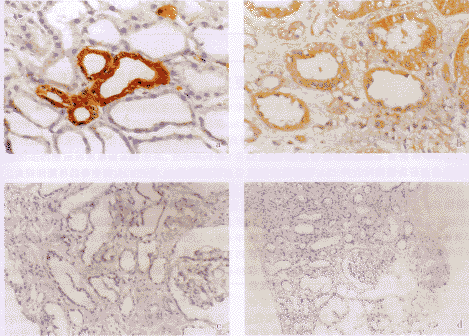
2.1 移植肾组织中CsA分布 (附图b)。本组11例肾移植功能减退患者中,有8例(72.7%)CsA阳性,对照组CsA为阴性。CsA阳性率在肾小管为72.7%(8/11),小血管为45.5%(5/11),肾小球为27.3%(3/11)(表2)。
, http://www.100md.com
2.2 P-gp在移植肾组织中表达 (附图a)。在肾小管表达为81.8%(9/11),小血管为27.2%(3/11),肾小球为18.1%(2/11)(表2)。
2.3 CsA和P-gp在移植肾组织中分布具有一致性,二者同时分布在肾小管最多,共7例(63.6%)。
2.4 患者血中环孢素浓度超过正常值[2]的次数,与CsA在移植肾组织中分布不一致。其中2例CsA阴性的患者,血中浓度曾经超过正常范围。肾移植手术时间的长短与移植肾组织中CsA和P-gp表达强度也无相关性(Spearman等级相关分析,r=0.17,P>0.05)。
2.5 对照组CsA在正常肾组织中表达均为阴性,P-gp表达分别为2例(+),1例(++),1例(-)。
, http://www.100md.com
Fig.1. Histochemical staining of CsA and P-gp in renal allograft biospy tissue sections
a.P-gp(×132) b.CsA(×132) c.P-gp control(×66) d.CsA control(×33)Table 2. The expression of CsA and P-gp in renal allograft Patientnumber
Months
after TX
CsA
P-gp
Times of increased
, 百拇医药
CsA detection
Tubules
Glomeruli
Vessels
Tubules
Glomeruli
Vessels
1
120
(++)
(-)
(++)
, 百拇医药
(++)
(-)
(+)
0
2
76
(+++)
(+)
(+++)
(+++)
(+)
(-)
7(359.2~632.2)
, 百拇医药
3
58
(-)
(-)
(-)
(+++)
(-)
(-)
1(429.3)
4
47
(-)
(-)
, 百拇医药
(-)
(-)
(-)
(-)
3(402.9~476.3)
5
48
NA
NA
NA
(++)
(-)
(-)
, 百拇医药
2(339.3~356.0)
6
38
(++)
(-)
(+)
(++)
(-)
(+)
4(394.8~867.9)
7
31
(+++)
, http://www.100md.com
(+++)
(+++)
(-)
(-)
(-)
4(305.0~379.8)
8
29
(++)
(-)
(++)
(++)
(+)
, 百拇医药
(-)
0
9
9
(+++)
(++)
(-)
(+)
(-)
(-)
3(377.4~487.7)
10
4
, 百拇医药
(+~++)
(-)
(-)
(+++)
(-)
(+++)
7(315.7~438.2)
11
0.5
(+)
(-)
(-)
(+)
, 百拇医药
(-)
(-)
1(504.9)
TX:transplantation;NA:data not available3 讨 论
环孢素的应用使人/肾存活率有了显著提高,但CsA肾毒性却成为移植肾功能减退的重要因素之一。肾移植术后肾功能减退通常由急性排斥、慢性排斥、CsA导致的肾毒性以及其它肾脏疾病引起[3,4],临床上若急性排斥合并CsA肾毒性或慢性排斥合并慢性CsA肾中毒时则难以鉴别。
急性CsA中毒的诊断可通过监测血中浓度加以判定,但CsA慢性肾毒性的诊断则比较困难,因为15%患者的CsA血浓度在正常范围[5],因此常需借助于病理检查。CsA导致肾脏出现葱皮样变(或片状纤维化)和玻璃样动脉粥样硬化的病理改变[6],在肾小管上皮出现空泡样变性,有时在线粒体内出现粥样小滴。本组7/11例(63.6%)患者HE切片中,出现不同程度肾小管上皮空泡样变性、玻璃样变和肾小管萎缩等病理改变。这些变化说明CsA已对移植肾上皮细胞造成了损害[7,8]。然而,这些病理变化缺乏特异性,因此,很难单纯依赖普通的HE染色方法将其与排斥反应鉴别[1]。
, 百拇医药
因此,我们利用免疫组织化学的方法在组织中直接定位检测CsA,以探讨CsA肾毒性在移植肾功能减退中的作用,为临床调整环孢素的使用提供参考。本组11例移植肾功能减退患者中,9例明确诊断为慢性排斥(81.8%),其中6例(66.7%)检测到CsA。即这6例慢性排斥患者可能同时合并有慢性CsA肾中毒。另外2例CsA阳性患者中,一例诊断为早期急性排斥,CsA血浓度超过正常值仅1次,经调整免疫抑制药物移植肾功能恢复正常;另一例病理检查排除排斥反应,但出现肾小管上皮变性、腔隙扩张,间质毛细血管扩张、充血等CsA慢性肾毒性的病理改变,而且CsA血浓度在短期内多次超过正常范围,因此,慢性CsA肾中毒的可能性较大。
Jette等[1]报道CsA能够诱导多药耐药蛋白(P-gp)的表达增加,而对肿瘤细胞CsA则能够阻止细胞P-gp表达,增强肿瘤细胞对化疗药物的耐药性。因此,我们检测了本组病例肾穿组织P-gp表达的情况。结果显示,本组81.8%(9/11)的病例有P-gp表达,同时有P-gp和CsA表达的病例占63.6%(7/11例)。这可能是因为CsA作用于移植肾组织,诱导P-gp的表达增加,从而发挥P-gp蛋白“泵”的功能,以将环孢素排出细胞外而达到对细胞解毒的作用[1]。这一现象提示,通过监测移植肾组织中P-gp的表达,可以间接反应CsA肾毒性的情况。
, 百拇医药
CsA的相对不溶性使其易于沉积在肾小管上皮细胞中,这是它最易导致肾小管上皮细胞损伤的重要原因[5]。本组病例中,CsA和P-gp在肾小管表达最多,进一步提示CsA可能主要沉积在肾小管上皮导致肾毒性。
检测移植肾组织中CsA和P-gp表达,为寻找诊断CsA导致肾毒性的方法提供了线索。为证实上述结论的正确性,除需进一步增加病例外,尚需对应用CsA而移植肾功能正常的移植受者肾组织进行对照观察研究。
倪灿荣(上海第二军医大学免疫病理室)
唐孝达(上海市第一人民医院泌尿科 上海,200080)
参考文献
1,Jette L,Beaulieu E,Leclerc JM et al.Cyclosporine A treatment induces over expression of P-glycoprotein in the kidney and other tissues.Am J Physiol,1996 May,270(5 Pt 2):F756
, 百拇医药
2,朱有华,孟 钢,闵志廉等.肾移植受者环孢素A治疗窗浓度的临床研究.中华泌尿外科杂志,1998,2:67
3,Benigni A,Bruzzi I,Mister M et al.Nature and mediators of renal lesions in kidney transplant patients given cyclosporine for more than one year.Kidney Int,1999 Feb,55(2):674
4,Al-Awwa IA,Hariharan S,First MR.Importance of allograft biopsy in renal transplant recipients:correlation between clinical and histological diagnosis.Am J Kidney Dis,1998 Jun,31(6 Suppl 1):S15
, 百拇医药
5,郑绮宜,郑克立.移植与环孢素.见:苏泽轩,于立新,黄洁夫主编.现代移植学.北京:人民卫生出版社,1998.170
6,Mihastsh MJ,Antonovych T,Bohman S-O et al.Cyclosporine A nephropathy:standardization of the evaluation of kidney biopsies.Clin Nephrol,1994,41:23
7,Mihastsh MJ,Thiel G,Ryffel B.Histopathology of cyclosporin nephrotoxicity.Transplant Proc,1988,20:759
8,Solez K,Axelsen RA,Benediktsson H et al.International standardization of criteria for the histologic diagnosis of renal allograft rejection:The Banff working classification of kidney transplant pathology.Kidney Int,1993,44:411
收稿1999-12-10
修回1999-12-25, http://www.100md.com
单位:凡杰(上海市第一人民医院泌尿科 上海,200080);王祥慧(上海市第一人民医院泌尿科 上海,200080);徐达(上海市第一人民医院泌尿科 上海,200080);王跃闽(上海市第一人民医院泌尿科 上海,200080);肖家全(上海市第一人民医院泌尿科 上海,200080);刘永(上海市第一人民医院泌尿科 上海,200080)
关键词:肾移植;免疫组织化学;环孢素
肾脏病与透析肾移植杂志000106 摘 要 目的:探讨免疫组织化学EnVision方法在移植肾功能减退患者肾组织中检测环孢素A(CsA)的可能性及其意义。 方法:收集我院1988年至1998年间肾移植术后长期服用CsA、血肌酐增高,临床表现为移植肾功能减退的患者11例,在B超引导下从移植肾上极、下极穿刺活检,正常肾组织作为对照组。采用免疫组织化学染色技术检测CsA及多药耐药P-gp蛋白在移植肾组织中的表达,观察其在组织切片中的形态学变化。 结果:本组11例肾移植患者中,有8例(72.7%)CsA阳性,对照组CsA为阴性;CsA以肾小管阳性率最高(72.7%),小血管次之(45.5%),肾小球最低(27.3%)。P-gp在移植肾组织中也以肾小管表达最高(81.8%),小血管次之(27.2%),肾小球最低(18.1%)。CsA和P-gp在移植肾组织中表达具有一致性,二者同时阳性以肾小管最高,共7例(63.6%)。患者血中环孢素浓度超过正常值的次数,与环孢素在移植肾组织中的阳性率不一致。肾移植后时间的长短与移植肾组织中CsA和P-gp表达程度不相关。 结论:检测移植肾肾组织中CsA的表达,有可能成为肾移植术后监测CsA肾毒性的一个重要指标,而同时检测多药耐药P-gp蛋白在移植肾组织中的表达,则可以提高诊断准确性。
, http://www.100md.com
CLINICAL SIGNIFICANCE OF CYCLOSPORINE A DETECTION IN RENAL ALLOGRAFTS
FAN Jie WANG Xianghui XU Da WANG Yaomin XIAO Jiaquan LIU Yong NI Canrong TANG Xiaoda
(Department of Urology,Shanghai First Peoples Hospital,Shanghai,200080)
OBJECTIVE To study the relationship between cyclosporine A nephrotoxicity and renal allograft dysfunction by detecting cyclosporine A(CsA)in renal allografts. METHODOLOGY Eleven patients on triple immunosuppressive therapy(CsA,axathioprien and steroid)with renal graft dysfunction after transplantation(increased serum creatinine>190 μmol/L)underwent allograft biopsy.Under the guidance of B mode ultrasound,a protocol biopsy of the renal graft was performed and autopsy tissue sections from normal kidney served as control group.The expressions of cyclosporine A and P-gp in renal allografts were detected by using immunohistochemical staining technique in 9 cases of chronic rejection,1 case of early acute rejection,and 1 case having no lesion in histological examination.Light microscopy examination and localization of CsA and P-gh in the graft specimen were evaluated with respect to the patterns of histologic lesion. RESULTS The expression of CsA was positive in eight out of 11 renal transplant recipients with allograft dysfunction,and was all negative in the control group.The incidence of the expression of CsA was 72.7%(8/11)in renal tubule,45.5%(5/11)in arteriol and 27.3%(3/11)in glomeruli respectively.The expression of P-gp in renal tubule was 81.8%(9/11),in arteriol 27.2%(3/11)and in glomeruli 18.1%(2/11)respectively.The incidence of the expression of CsA and P-gp(predominantly in renal tubule)was 63.6%(7/11).7 of 11 renal transplant recipients who underwent the protocol biopsy had CsA nephrotoxicity proved by histological examination. CONCLUSION Combined detection of cyclosporine A and P-gp is of clinical significance in the diagnosis of cyclosporine A nephrotoxicity.
, 百拇医药
Key words kidney transplantation immunohistochemistry cyclosporine A
环孢素A(CsA)肾毒性是急、慢性移植肾功能减退的原因之一,但CsA肾毒性常难以与其它影响移植肾功能减退的危险因素相鉴别。我们利用免疫组织化学的方法研究CsA在肾功能减退患者移植肾组织中的分布,以探索利用免疫组织化学方法在移植肾组织中检测CsA的可能性,为诊断CsA肾毒性提供新的方法。另外,CsA能够诱导肾脏等组织细胞多药耐药蛋白(P-gp)的表达增加[1],因此联合检测移植肾组织中CsA、P-gp的表达,则有可能提高诊断CsA肾毒性的准确性。
1 对象和方法
1.1 临床资料 我院1988年至1998年间肾移植患者11例,临床表现为移植肾功能减退,其临床资料见表1,所有病例均在我院接受长期随访。以4例肾癌手术切除后的肾组织无癌区,(病理证实为正常肾组织),作为对照组。对照组活检取材大小为0.4cm×0.6cm×0.7cm。实验组分别在B超引导下从移植肾上极、下极穿刺活检,组织块大小约为1.50cm×0.05cm×0.01cm,每例取2~3个组织块。活检组织用100 g/L的甲醛溶液固定,脱水,石蜡包埋,同时作HE染色。
, http://www.100md.com
1.2 试剂
1.2.1 P-gp混合单抗 JSB,C219,C494为丹麦DAKO公司产品,1∶50稀释。
1.2.2 环孢素A单抗 瑞士Norvatis公司产品。
1.2.3 其它试剂 EnVision Kit为丹麦DAKO公司产品。
1.3 实验方法 石腊切片常规脱腊,PBS缓冲液冲洗,微波抗原修复,然后用3%H2O2阻断内源性过氧化酶,PBS洗3×3min,依次加入一抗(CsA单抗或P-gp混合单抗),4℃过夜,PBS洗3×3min,再滴加EnVision多聚物酶复合物孵育37℃ 30min。0.05%DAB+0.03%H2O2显色,苏木素复染,逐级脱水,透明封片,光镜观察。用PBS代替一抗作阴性对照,HE染色明确病理诊断。
, 百拇医药
1.4 免疫组织化学染色结果判定
1.4.1 P-gp表达 ①:50%以上的肾小管、肾小球细胞及小血管呈阳性表达;②:25%~50%的肾小管、肾小球细胞及小血管呈阳性表达;③+:25%以下的肾小管、肾小球细胞及小血管呈阳性表达;④正常(-):内散在个别阳性细胞,一般少于10%。
1.4.2 CsA表达 ①:50%以上的肾小管、肾小球细胞及小血管阳性;②:25%~50%的肾小管、肾小球细胞及小血管阳性;③+:25%以下的肾小管、肾小球细胞及小血管阳性;④正常(-):内散在个别阳性细胞,一般少于10%。
Table 1. The relevant clinical data of the 11 recipients with graft dysfunction Patientnum
Patient
, 百拇医药
Age
TX
Date
CMV
IgG
Increased
serum lipids
serum
Cr(μmol/L)
proteinuria
Doppler US
Pathology
, http://www.100md.com
UT obstruction
RI
1
44
88-12-13
(+)
↑
480
(++)
(-)
Normal
CR
2
, 百拇医药
28
92-04-30
(-)
↑
580
(+)
(-)
Normal
CR
3
34
93-10-01
(-)
, http://www.100md.com
↑
300
(-)
(-)
Normal
CR
4
41
94-04-26
(-)
↑
250
(-)
, 百拇医药
(-)
Normal
CR
5
20
94-07-29
(+)
↑
370
(+)
(-)
Normal
CR
, 百拇医药
6
40
95-06-01
(-)
↑
240
(+++)
(-)
Normal
CR
7
28
95-12-06
, 百拇医药
(-)
↑
190
(+-)
(-)
Normal
CR
8
24
96-07-06
(+)
↑
340
, http://www.100md.com
(++++)
(-)
Normal
CR
9
29
97-09-27
(-)
↑
360
(-)
(-)
Normal
, 百拇医药
CR
10
38
98-09-01
(-)
↑
280
(-)
(-)
Normal
No rejection
11
28
, http://www.100md.com
98-09-23
(-)
↑
270
(++++)
(-)
Normal
Early AR
2 结 果
2.1 移植肾组织中CsA分布 (附图b)。本组11例肾移植功能减退患者中,有8例(72.7%)CsA阳性,对照组CsA为阴性。CsA阳性率在肾小管为72.7%(8/11),小血管为45.5%(5/11),肾小球为27.3%(3/11)(表2)。
, http://www.100md.com
2.2 P-gp在移植肾组织中表达 (附图a)。在肾小管表达为81.8%(9/11),小血管为27.2%(3/11),肾小球为18.1%(2/11)(表2)。
2.3 CsA和P-gp在移植肾组织中分布具有一致性,二者同时分布在肾小管最多,共7例(63.6%)。
2.4 患者血中环孢素浓度超过正常值[2]的次数,与CsA在移植肾组织中分布不一致。其中2例CsA阴性的患者,血中浓度曾经超过正常范围。肾移植手术时间的长短与移植肾组织中CsA和P-gp表达强度也无相关性(Spearman等级相关分析,r=0.17,P>0.05)。
2.5 对照组CsA在正常肾组织中表达均为阴性,P-gp表达分别为2例(+),1例(++),1例(-)。

, http://www.100md.com
Fig.1. Histochemical staining of CsA and P-gp in renal allograft biospy tissue sections
a.P-gp(×132) b.CsA(×132) c.P-gp control(×66) d.CsA control(×33)Table 2. The expression of CsA and P-gp in renal allograft Patientnumber
Months
after TX
CsA
P-gp
Times of increased
, 百拇医药
CsA detection
Tubules
Glomeruli
Vessels
Tubules
Glomeruli
Vessels
1
120
(++)
(-)
(++)
, 百拇医药
(++)
(-)
(+)
0
2
76
(+++)
(+)
(+++)
(+++)
(+)
(-)
7(359.2~632.2)
, 百拇医药
3
58
(-)
(-)
(-)
(+++)
(-)
(-)
1(429.3)
4
47
(-)
(-)
, 百拇医药
(-)
(-)
(-)
(-)
3(402.9~476.3)
5
48
NA
NA
NA
(++)
(-)
(-)
, 百拇医药
2(339.3~356.0)
6
38
(++)
(-)
(+)
(++)
(-)
(+)
4(394.8~867.9)
7
31
(+++)
, http://www.100md.com
(+++)
(+++)
(-)
(-)
(-)
4(305.0~379.8)
8
29
(++)
(-)
(++)
(++)
(+)
, 百拇医药
(-)
0
9
9
(+++)
(++)
(-)
(+)
(-)
(-)
3(377.4~487.7)
10
4
, 百拇医药
(+~++)
(-)
(-)
(+++)
(-)
(+++)
7(315.7~438.2)
11
0.5
(+)
(-)
(-)
(+)
, 百拇医药
(-)
(-)
1(504.9)
TX:transplantation;NA:data not available3 讨 论
环孢素的应用使人/肾存活率有了显著提高,但CsA肾毒性却成为移植肾功能减退的重要因素之一。肾移植术后肾功能减退通常由急性排斥、慢性排斥、CsA导致的肾毒性以及其它肾脏疾病引起[3,4],临床上若急性排斥合并CsA肾毒性或慢性排斥合并慢性CsA肾中毒时则难以鉴别。
急性CsA中毒的诊断可通过监测血中浓度加以判定,但CsA慢性肾毒性的诊断则比较困难,因为15%患者的CsA血浓度在正常范围[5],因此常需借助于病理检查。CsA导致肾脏出现葱皮样变(或片状纤维化)和玻璃样动脉粥样硬化的病理改变[6],在肾小管上皮出现空泡样变性,有时在线粒体内出现粥样小滴。本组7/11例(63.6%)患者HE切片中,出现不同程度肾小管上皮空泡样变性、玻璃样变和肾小管萎缩等病理改变。这些变化说明CsA已对移植肾上皮细胞造成了损害[7,8]。然而,这些病理变化缺乏特异性,因此,很难单纯依赖普通的HE染色方法将其与排斥反应鉴别[1]。
, 百拇医药
因此,我们利用免疫组织化学的方法在组织中直接定位检测CsA,以探讨CsA肾毒性在移植肾功能减退中的作用,为临床调整环孢素的使用提供参考。本组11例移植肾功能减退患者中,9例明确诊断为慢性排斥(81.8%),其中6例(66.7%)检测到CsA。即这6例慢性排斥患者可能同时合并有慢性CsA肾中毒。另外2例CsA阳性患者中,一例诊断为早期急性排斥,CsA血浓度超过正常值仅1次,经调整免疫抑制药物移植肾功能恢复正常;另一例病理检查排除排斥反应,但出现肾小管上皮变性、腔隙扩张,间质毛细血管扩张、充血等CsA慢性肾毒性的病理改变,而且CsA血浓度在短期内多次超过正常范围,因此,慢性CsA肾中毒的可能性较大。
Jette等[1]报道CsA能够诱导多药耐药蛋白(P-gp)的表达增加,而对肿瘤细胞CsA则能够阻止细胞P-gp表达,增强肿瘤细胞对化疗药物的耐药性。因此,我们检测了本组病例肾穿组织P-gp表达的情况。结果显示,本组81.8%(9/11)的病例有P-gp表达,同时有P-gp和CsA表达的病例占63.6%(7/11例)。这可能是因为CsA作用于移植肾组织,诱导P-gp的表达增加,从而发挥P-gp蛋白“泵”的功能,以将环孢素排出细胞外而达到对细胞解毒的作用[1]。这一现象提示,通过监测移植肾组织中P-gp的表达,可以间接反应CsA肾毒性的情况。
, 百拇医药
CsA的相对不溶性使其易于沉积在肾小管上皮细胞中,这是它最易导致肾小管上皮细胞损伤的重要原因[5]。本组病例中,CsA和P-gp在肾小管表达最多,进一步提示CsA可能主要沉积在肾小管上皮导致肾毒性。
检测移植肾组织中CsA和P-gp表达,为寻找诊断CsA导致肾毒性的方法提供了线索。为证实上述结论的正确性,除需进一步增加病例外,尚需对应用CsA而移植肾功能正常的移植受者肾组织进行对照观察研究。
倪灿荣(上海第二军医大学免疫病理室)
唐孝达(上海市第一人民医院泌尿科 上海,200080)
参考文献
1,Jette L,Beaulieu E,Leclerc JM et al.Cyclosporine A treatment induces over expression of P-glycoprotein in the kidney and other tissues.Am J Physiol,1996 May,270(5 Pt 2):F756
, 百拇医药
2,朱有华,孟 钢,闵志廉等.肾移植受者环孢素A治疗窗浓度的临床研究.中华泌尿外科杂志,1998,2:67
3,Benigni A,Bruzzi I,Mister M et al.Nature and mediators of renal lesions in kidney transplant patients given cyclosporine for more than one year.Kidney Int,1999 Feb,55(2):674
4,Al-Awwa IA,Hariharan S,First MR.Importance of allograft biopsy in renal transplant recipients:correlation between clinical and histological diagnosis.Am J Kidney Dis,1998 Jun,31(6 Suppl 1):S15
, 百拇医药
5,郑绮宜,郑克立.移植与环孢素.见:苏泽轩,于立新,黄洁夫主编.现代移植学.北京:人民卫生出版社,1998.170
6,Mihastsh MJ,Antonovych T,Bohman S-O et al.Cyclosporine A nephropathy:standardization of the evaluation of kidney biopsies.Clin Nephrol,1994,41:23
7,Mihastsh MJ,Thiel G,Ryffel B.Histopathology of cyclosporin nephrotoxicity.Transplant Proc,1988,20:759
8,Solez K,Axelsen RA,Benediktsson H et al.International standardization of criteria for the histologic diagnosis of renal allograft rejection:The Banff working classification of kidney transplant pathology.Kidney Int,1993,44:411
收稿1999-12-10
修回1999-12-25, http://www.100md.com