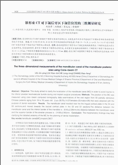
锥形束CT对下颌后牙区下颌管位置的三维测量研究(2)
 |
| 第1页 |
参见附件。
目前较为通用的研究下颌管走行的影像学方法为拍摄CBCT和多层螺旋CT,关于下颌管的精细解剖研究同影像学检查结果是一致的[6-8]。在种植手术中,因为下颌管在骨内的走行轨迹存在规律的变化,如果能准确的测量可用牙槽嵴的高度,在骨量不足的情况下也有可能避免复杂且昂贵的植骨手术。而常规的根尖片以及全景片则因为其二维图像的特点不能反映下颌管的三维位置关系。
本研究旨在通过CBCT检查评价我院所在地区人群下颌骨解剖学形态以及下颌管三维位置的统计学指标,是初期的小样本研究,笔者将继续统计信息,积累大样本资料。
4 结论
通过CBCT测量得到下颌管及下颌骨的形态学数据,对种植术前确定下颌管的解剖位置有重要的临床指导意义,且大部分牙列缺损患者均具有种植修复缺牙的骨量条件。
[参考文献]
[1]de Oliveira Junior,M. R.,Saud, A.L.,Fonseca, D. R.,et al.Morphometrical analysis of the human mandibular canal:a CT investigation[J].Surg Radiol Anat,2010,DOI 10.1007/s00276-010-0708-3.
[2]Yu IH,Wong YK.Evaluation of mandibular anatomy related to sagittal split ramus osteotomy using 3-dimensional computed tomography scan images[J].Int J Oral Maxillofac Surg,2008,37:521-528.
[3]Kriwalsky MS,Maurer P,Veras RB,et al.Risk factors for a bad split during sagittal split osteotomy[J].Br J Oral Maxillofac Surg,2008,46:177-179.
[4]Trauner R,Obwegeser HL.The surgical correction of mandibular prognathism and retrognathia with consideration of genioplasty.Part I.Surgical procedures to correct mandibular prognathism and reshaping of the chin[J].Oral Surg Oral Med Oral Pathol,1957,10:677-689.
[5]钱文涛,樊林峰,徐光宙,等.CBCT观察影像重叠的下颌第三磨牙与下颌管的位置关系[J].口腔颌面外科杂志,2010,20(6):398-403.
[6]Naitoh M,Nakahara K,Suenaga Y,et al.Comparison between cone-beam and multislice computed tomography depicting mandibular neurovascular canal structures[J].Oral Surg Oral Med Oral Pathol Oral Radiol Endod,2010,109:e25-e31.
[7]吴银洲,吴利民,胡圣望.100例下颌骨下颌管形态及其至牙槽嵴距离的观测研究[J].口腔颌面修复学杂志,2004,5(3):180-181.
[8]王 蓓,房洪波,徐 兵,等.100例正常人下颌管的三维测量[J].实用口腔医学杂志,2010,26(2):223-226.
[收稿日期]2012-05-22 [修回日期]2012-07-16
编辑/何志斌
您现在查看是摘要介绍页,详见PDF附件(2214kb)。