补肾丹对衰老模型大鼠脑海马组织P53基因表达的影响(2)
 |
| 第1页 |
参见附件(2698KB,2页)。
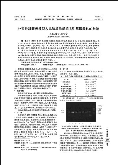
本实验应用RT-PCR和Western Blot的方法,检测补肾丹对D-半乳糖所致亚急性衰老模型大鼠脑海马组织P53基因表达的影响。结果表明,衰老模型大鼠的P53基因表达明显增高,与空白组比较有统计学意义(P<0.05),补肾丹各剂量组相对模型组而言,有明显的降低,其中补肾丹中剂量组效果最好,说明补肾丹能够通过降低P53基因的过度表达来达到延缓脑细胞衰老的目的。
参考文献
[1] 刘学丽,朱延勤,潘伟娜,等.对人鼠半乳糖性自内障实验模型的探讨[J].北京实验动物科学与管理,1994,11(2):2-3.
[2] Kuan Nk, Passaro E Jr.Apoptosis: programmed cell death[J].Arch Surg, 1998, 133 (7):773-775.
[3] Yand E, Korameyer SJ. Molecular mechanis of apoptosis: a discourse on the bcl-2 family and cell death Blood[J]. 1996: 88(2):386.
[4] Uren AG, Vaux DL.Molecular and clinical aspects of apoptosis[J]. Pharmacol Ther,1996, 72(1):37.
[5] Levine A J. p53, the cellular gatekeeper for growth and division[J]. Cell,1997,88:323-331.
[6] KoL J.Prives C.p53:puzz leand paradigm[J].Genes & Development,1996,10:1054-1072.
[7] Gottlieb T M,Oren M.P53 in growth control and neoplasia[J]. Biochimica et Biophysics Acta,1996,12:77.
[8] El-Deiry W S. Regulation of p53 downstream genes[J]. Seminars in Cancer Biology, 1998, 8:345-357.
[9] Smith J R, Pereira-Smith O M. Replicative senescence: Implications for in vivo aging and tumor suppression[J]. Science, 1996, 273: 63-66.
[10] Chin L, Artand S E, Shen Q, et al ......
您现在查看是摘要介绍页,详见PDF附件(2698KB,2页)。