中枢神经细胞瘤的CT诊断(1)
 |
| 第1页 |
参见附件(1185KB,2页)。
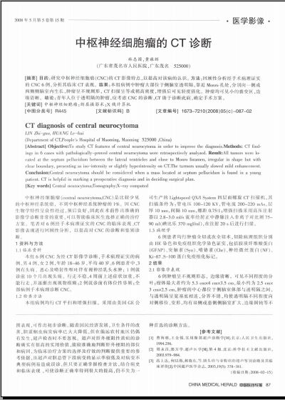
[摘要] 目的:研究中枢神经细胞瘤(CNC)的CT影像特点,以提高对该病的认识。方法:回顾性分析经手术病理证实的CNC 6例,分析其临床CT表现。结果:本组病例中肿瘤大部位于侧脑室透明隔,靠近Monro孔处,分别向一侧或两侧侧脑室内生长,肿瘤呈不规则形, CT扫描呈等或稍高密度,增强后可见轻度强化。肿瘤均可见小的囊变区,边缘清晰。结论:青年人位于透明隔的肿瘤,应考虑CNC的诊断,CT助于诊断此病,确定手术方案。
[关键词] 中枢神经细胞瘤;断层摄影术;X线计算机
[中图分类号]R445 [文献标识码]B [文章编号]1673-7210(2008)05(c)-087-02
CT diagnosis of central neurocytoma
LIN Zhi-guo, HUANG Lv-hui
(Department of CT,People's Hospital of Maoming, Maoming525000 ,China)
[Abstract] Objective:To study CT features of central neurocytoma in order to improve the diagnosis.Methods: CT findings in 6 cases with pathologically-proved central neurocytoma were retrospectively analyzed. Result:All tumors were located at the septum pellucidum between the lateral ventricles and close to Monro foramen, irregular in shape but with clear boundary, presenting as iso-intensity or slightly hyperintensity on CT ......
您现在查看是摘要介绍页,详见PDF附件(1185KB,2页)。