肾透明细胞癌与肾乳头状癌CT、MRI表现的比较(3)
 |
| 第1页 |
参见附件。
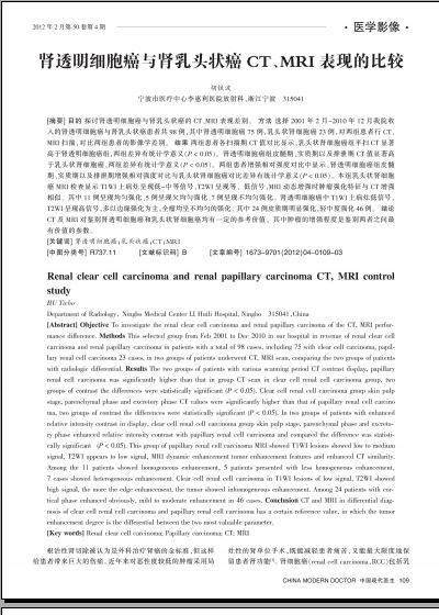
综上所述,CT及MRI对鉴别肾透明细胞癌和乳头状肾细胞癌均有一定的参考价值,其中肿瘤的增强程度是鉴别两者之间最有价值的参数。
[参考文献]
[1] Fornara P,Hoda MR. Renal cell carcinoma[J]. Urologe A,2011,50 (Suppl 1):219-222.
[2] Volpe A, Patard JJ. Prognostic factors in renal cell carcinoma[J]. World J Urol,2010,28(3):319,327.
[3] 吴晓华,贺文,马大庆,等. 乳头状肾细胞癌的螺旋CT诊断及鉴别诊断[J]. 中国医学影像技术,2009:25(6):1063-1065.
[4] Amin MB, Amin MB, Tamboli P, et al. Prognostic impact of histologic subtyping of adult renal epithelial neoplasms: an experience of 405 cases[J]. Am J Surg Pathol, 2002, 26(3): 281-291.
[5] Cheville JC, Lohse CM, Zineke H, et al. Comparisons of outcome and prognostic features among histologic subtypes of renal cell carcinoma[J]. Am J Surg Pathol, 2003, 27: 612-624.
[6] 项剑瑜,刘绪明,许加峻,等. 肾透明细胞癌的MRI诊断[J]. 医学影像学杂志2010:20(1):83-86.
[7] Fujimoto H, Wakao F, Moriyama N. Alveolar architecture of clear cell renal carcinomas (
[8] Sheir KZ,Mohamed E,Mosbah A,et al. Differentiation of renal cell carcinoma subypes by multislice computerized tomography[J]. J Urol,2005, 147:451-455.
[9] 张京刚,王希明,胡春洪,等. 乳头状肾细胞癌的CT表现[J]. 放射学实践,2011:26(6):627-630.
(收稿日期: 2011-11-24)
您现在查看是摘要介绍页,详见PDF附件(2958kb)。