大鼠非酒精性脂肪肝造模方法的改进
非酒精性脂肪肝;动物模型;丙基硫氧嘧啶;大鼠,王倩,管小琴,王倩,通讯作者:,Improvementofinductionmethodofnon-alcoholicfattylivermodelinrats,QianWang,Xiao-QinGuan
 |
| 第4页 |
 |
| 第1页 |
参见附件(970KB,6页)。
王倩, 管小琴, 重庆医科大学病理教研室 重庆市 400016
王倩, 医学硕士, 主要从事肝胆病理研究.
通讯作者: 管小琴, 400016, 重庆市渝中区医学院路1号, 重庆医科大学病理教研室. guanxiaoqin2003@yahoo.com.cn
收稿日期: 2006-12-27 接受日期: 2007-01-20
Improvement of induction method of non-alcoholic fatty liver model in rats
Qian Wang, Xiao-Qin Guan
Qian Wang, Xiao-Qin Guan,Department of Pathology, Chongqing University of Medical Sciences, Chongqing 400016, China
Correspondence to:Xiao-Qin Guan, Department of Pathology, Chongqing University of Medical Sciences, 1 Yuzhong Road, Yuzhong District, Chongqing 400016, China. guanxiaoqin2003@yahoo.com.cn
Received:2006-12-27 Accepted:2007-01-20
Abstract
AIM: To establish a rapid rat model of non-alcoholic fatty liver.
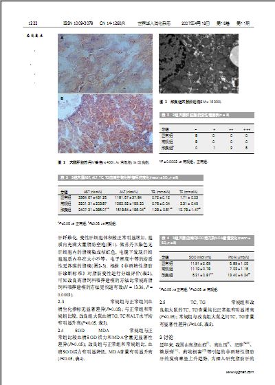
METHODS: Twenty-four male Sprague Dawley rats were averagely and randomly divide into group A, B and C, fed with normal diet, routine high-fat diet and routine high-fat diet plus sucrose, propylthiouracil, and sodium cholate, respectively. The general conditions and weight changes were dynamically observed for 5 weeks, and then all the rats were killed. The pathological changes of liver tissues were observed by HE staining, and Sudan IV staining and electron microscopy were used to investigate the presence status of cytoplasmic lipid droplets in liver cells. The following indexes were compared between the three groups, including serum levels of triglyceride (TG), total cholesterol (TC), alanine aminotransferase (ALT), aspartate aminotransferase (AST), malondialdehyde (MDA), and superoxide dismutase (SOD), and tissue contents of TG and TC.
RESULTS: Starting from the fourth week, the weights of rats were significantly decreased in group A and C as compared with those in group B (249.63 ± 34.25, 241.88 ± 20.75 vs 275.38 ± 6.59, P < 0.05), but there was no marked difference between group A and C (P > 0.05). In the fifth week, light microscopy showed a great number of fatty vacuoles in liver cells, and electron microscopy confirmed the presence of abundant lipid droplets ......
您现在查看是摘要介绍页,详见PDF附件(970KB,6页)。