Down综合征16三体小鼠胃的神经发育观察
作者:李继承 Busch LC Khnel W
单位:李继承 310006杭州,浙江大学医学院淋巴学研究室组织胚胎学教研室;Busch LC, Khnel W 德国Lübeck医科大学解剖学研究所
关键词:16三体鼠;胃;神经发育;蛋白基因产物9.5;免疫组织化学
Down综合征16三体小鼠胃的 神经发育观察 李继承 Busch LC K hnel W
【摘要】 目的 研究Down综合征动物模型16三体和正常同窝鼠支配胃的神经发育。方法 采用16三体鼠培育,同窝鼠胚胎龄(embryonic days, ED)13~18天细胞遗传学分析,蛋白基因产物9.5(protein gene product 9.5, PGP 9.5)免疫组化等方法对16三体小鼠胃的神经发育进行了研究。结果 正常同窝鼠,胎龄13天(ED13)来源于外胚层神经嵴的神经母细胞迁移并进驻胃壁;ED14神经元发出突起,形成原始神经网络;ED15形成简单排列的肌间神经丛,开始出现早期的神经节;ED16有分布规则的肌间神经丛;ED17神经母细胞进驻粘膜下层,形成粘膜下神经丛;ED18完整的胃神经丛形成,即粘膜下浅、深神经丛和肌间神经丛。与正常同窝鼠比较,16三体鼠胃神经系发育迟缓,ED14胃壁始有散在分布神经元。此后,胃神经系的发育与分化均较正常延迟,至ED18仅有肌间神经丛。根据胃神经系的发育程度和PGP9.5免疫反应强度作半定量分析及秩和检验,16三体鼠胃神经的发育明显落后于它们正常的同窝鼠,两者比较有显著性差异(P<0.05)。结论 16三体小鼠是公认的Down综合征动物模型,它除了有多系统和多器官的畸型外,还发现有小鼠胃神经丛发育迟缓,粘膜下神经丛缺失。
, 百拇医药
Observation on the development of stomach innervation in trisomy 16 mouse, an animal model for Down syndrome
LI Jicheng*, Busch LC, Khnel W.* Department of Lymphology, Department of Histology and Embryology, Zhejiang University Medical School, Hangzhou 310006 P.R.China.Email:lijc@mail.hz.zj.cn
【Abstract】 Objective To study the nervous development in the stomach of trisomy 16 mouse embryos and their normal littermates from embryonic days 13-18(ED 13-18) by using primary antibody against protein gene product (PGP) 9.5.Methods (A) trisomy 16 mouse breeding; (B) trisomy 16 mouse embryos were confirmed by examining their chromosomes;(C) PGP 9.5 immunohistochemical method.Results In normal littermates, the precursor cells from the neural crest migrated into stomach at ED13.Numerous nervous processes connected each other and formed early nervous networks at ED14. Original ganglia appeared in the muscular plexus with very simple arrangment at ED15.The developed myenteric and submucosal plexuses were found at ED16 and ED17 respectively.The myenteric plexus and the internal and external submucosal plexuses were differentiated at ED18.In comparison with the normal littermates the trisomy 16 mice's stomach nervous development was much slower. Their immature neurons did not appear in the stomach until ED14; the development and differentiation of stomach nervous plexuses were delayed. A clear link was noted between the development of stomach plexuses and the density of PGP 9.5 immunostaining. A semiquantitative analysis and rank sum test of the data showed that the trisomy 16 mouse embryos were markedly retarded in nervous development of stomach, compared with their normal littermates (P<0.05). Conclusion Trisomy 16 mouse, as an animal model for Down syndrome,has abnormality not only in several systems and organs but also in stomach innervation. This is the first report on delayed nervous development in the stomach of trisomy 16 mice.
, 百拇医药
【Key words】 Trisomy 16 mouse Stomach Nervous development Protein gene product 9.5 Immunohistochemistry
Down综合征动物模型16三体小鼠常伴发多系统多器官的畸型。由于严重心血管系的发育障碍,16三体小鼠出生即不能成活。为此,学者们均以16三体胎鼠和它们正常同窝鼠作为研究Down综合征的动物模型。
Down综合征表现为先天愚型和多器官畸型,是人类首先报道的染色体疾病。Yoon等[1]统计,有95%的Down综合征是由21三体所致,其发生率约为1/700~1/1500。近年来,由于化学药物的滥用、病毒感染、杀虫剂广泛使用、放射线照射和母亲高龄等诸多因素,致使Down综合征发生率有上升趋势。已引起国际学术界的广泛关注,并加强了对Down综合征发病预防和16三体小鼠研究。
, http://www.100md.com
我们参加了德国Down综合征动物模型16三体小鼠的研究,并承担了16三体小鼠胃肠道神经系统发育的研究项目。我们采用蛋白基因产物(protein gene product,PGP) 9.5研究胃神经丛的发生和发育,首次报道了16三体小鼠胃肠道神经系的发育障碍。有关16三体小鼠肠神经系的研究已作另文报道。
1 材料与方法
1.1 PGP 9.5分子量2.45kb,由212个氨基酸组成。特异性地存在于神经元和神经内分泌细胞胞浆的羧基末端水解酶。PGP 9.5具有灵敏的神经定位表达作用。PGP 9.5的稀释液为含10%正常羊血清、0.01%叠氮钠和0.05%硫汞撒的PBS液(pH7.4)。
1.2 16三体小鼠繁殖和饲养 由NMR-1雌性小鼠与携带有两个罗伯逊易位的Rb(16,17),Rb(11,16)8LubtWLub3雄性小鼠交配繁殖16三体小鼠。并以观察到母鼠阴道粘液栓为Embryonic day(ED)1。
, 百拇医药
1.3 细胞遗传学分析
1.3.1 腹腔内注射秋水仙素(0.02mg/25~35mg体重)1小时后,以断颈法处死孕鼠,剖腹取胚胎。
1.3.2 从每个胚胎中取部分肝组织,经0.563%KCl低渗处理,甲醛∶乙醇(3∶1)固定,风干涂片,光镜观察。以镜下观察到两条Rb染色体和合计41条染色体为16三体鼠。
1.4 免疫组织化学方法 取16三体和正常同窝鼠固定在含4%多聚甲醛和2%苦味酸的磷酸缓冲液中2小时。取胃,在解剖镜下制备胃铺片,并作冰冻切片,切片厚度为12μm。按ABC和PAP免疫组化法,用4-氯-1萘酚为生色剂显色。所用试剂、工作浓度和作用时间见表1。并用PBS取代PGP9.5抗体作阴性对照。标本用Zeiss显微镜观察并拍照。
表1 试剂来源、工作浓度和作用时间
, 百拇医药
Tab 1 Reagent source, working concentration and reactive time
Reagent
(试剂)
Source
(来源)
Working
concentration
(工作浓度)
Reactive
time(h)
(作用时间)
, http://www.100md.com
(小时)
Rabbit anti-human
Ultraclone limited
PGP 9.5 antibodies
UK
1∶400
24
Goat anti-rabbit
DAKO Denmark
1∶100
12
Immunoglobulins
, 百拇医药
rabbit PAP
DAKO Denmark
1∶100
12
Goat anti-rabbit IgG
Siagma USA
1∶100
6
biotin conjugate
Sigma USA
1∶20
6
, 百拇医药
streptavidin
biotinylate
horseradish
peroxidase complex
1.5 胃神经丛发育的半定量统计分析 根据神经发育的形态特征和PGP9.5免疫反应强度,制定以下标准:无神经元及免疫反应阳性神经纤维(-);散在神经元,PGP9.5弱阳性(+);神经元发出突起,相邻神经元的突起相连,PGP9.5阳性(++);肌间神经丛和神经节形成,PGP9.5中等阳性(+++);粘膜下神经丛出现,肌间神经丛网络形成,PGP9.5强阳性(++++);粘膜下神经浅、深丛分化,高度复杂肌间神经丛网络形成(+++++)。秩和检验,统计分析。
2 结果
Down综合征动物模型16三体小鼠有严重的先天性心血管畸形,如室间隔大面积缺损,故出生后均死亡。根据我们观察,从ED13至ED18,16三体鼠存活率由36.4%降至9.3%。自ED15以后,16三体胎鼠多有严重的全身性水肿和发育不良,但取材时均需作细胞染色体检查,以发现有两条Rb染色体和合计41条染色体,才能确定16三体小鼠。
, http://www.100md.com
在ED13正常同窝鼠胃壁肌层,可观察到呈散在分布的来源于外胚层神经嵴的神经母细胞。16三体鼠则在ED14才能观察到PGP9.5阳性神经元。从神经母细胞进驻胃壁的发育时相上,16三体小鼠落后于它们的正常同窝鼠1天(图1a)。正常同窝鼠ED14胚胎龄期神经元已发出许多突起,形成原始的神经网络(图1b);ED15出现简单排列的神经丛和早期的神经节;ED16形成肌间神经丛,并可见分布规则的神经网络。16三体小鼠胃神经发育显著迟缓,ED15始见有PGP9.5弱阳性的神经突起,ED16的神经丛发育低下,神经纤维稀少。在冰冻切片PGP9.5免疫反应上,16三体鼠胃神经丛比它们同龄同窝鼠染色浅(图2)。ED17~18胎龄期,正常小鼠的胃壁神经丛分化为肌间神经丛和粘膜下神经丛。肌间神经丛由初级、次级神经索和更细小的神经纤维组成,沿初级神经索有许多发育良好的神经节分布。粘膜下神经丛是不规则的神经网络,由细小的神经纤维和少量散在分布的神经节组成,参与构成神经节的神经元数量少,PGP9.5强阳性神经元亦较少见。在ED18期,正常同窝鼠胃壁强阳性的神经元数量明显多于16三体鼠(图3)。同期的16三体小鼠仅见肌间神经丛,且神经网络稀疏、染色较浅,强阳性神经元少见,神经节无论在形态和数量上均少于正常小鼠。此外,三体小鼠另一个重要特征是它们的粘膜下神经丛发育受阻,胃粘膜下层无神经丛。
, 百拇医药
图1 用组织铺片法显示胃神经 a:ED14 16三体小鼠胃壁出现散在分布的神经元(↑);b:ED14正常同窝鼠胃神经网络神经元(↑);×250
Fig 1 The stomach innervation was observed by using whole mount preparation a:some neurons(arrows) with a scattering distribution appeared in stomach of ED 14 trisomy 16 mouse embryo;b:the nervous network of stomach in ED 14 normal littermate. Neurons (arrows) in the nervous network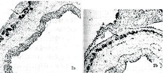
, http://www.100md.com
图2用切片法显示ED15 16三体和它们正常同窝鼠胃肌间神经丛,神经节(↑) a:正常同窝鼠胃神经;b:16三体小鼠胃神经,×100 Fig 2 Stomach myenteric plexus of ED15 trisomy 16 mouse embryo and its normal littermate by using sectioning a:normal littermate;b:trisomy 16 mouse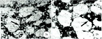
图3 用组织铺片法显示ED18 16三体和正常同窝鼠胃肌间神经丛、神经节(↑) a:16三体小鼠胃肌间神经丛;b:正常同窝鼠胃肌间神经丛;×250Fig 3 Stomach myenteric plexus of ED18 trisomy 16 and its normal littermate by using whole mount preparation a:trisomy 16 mouse;b:normal littermate
, 百拇医药
PGP9.5是一种神经元羧基末端水解酶,对神经元和神经纤维有很强的表达作用,其免疫反应的强度随胃神经丛发育而逐渐增强。根据胃神经丛发育和PGP9.5免疫反应强度,我们对16三体和它们正常同窝鼠进行了半定量分析及秩和检验(表2),比较同龄正常与16三体小鼠肌间神经丛和粘膜下神经丛的发育,发现在同一发育时相,16三体与正常同窝鼠的胃神经丛发育有明显的区别(P<0.05),前者胃神经发育迟缓(图4)。
表2 按时间顺序16三体和它们正常同窝鼠胃神经的胚龄发育情况
Tab 2 The development of the stomach innervation of trisomy 16 mouse embryos and their normal littermates in chronological sequence
ED18
, http://www.100md.com
ED17
ED16
ED15
ED14
ED13
Normal
mouse
(正常鼠)
+++++
++++
+++
+++
++
, 百拇医药
+
Trisomy 16*
mouse
(16三体鼠)
+++
+++
++
++
+
-
*P<0.05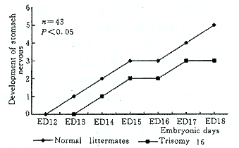
, http://www.100md.com
图4 16三体和正常同窝鼠胚胎龄和胃神经丛的发育
Fig 4 The devlopment of stomach nervous plexuses in trisomy 16 mouse embryos and their normal littermates
3 讨论
Down综合征是人类21染色体畸变引起的遗传病,又称为先天愚型,占所有智力迟滞患者的10%~15%。由于对Down综合征胃神经发育的直接研究尚有很大的困难,为此,我们采用16三体小鼠作为研究对象。16三体小鼠是由自然条件下罗伯逊易位小鼠的培养,所产生的非整倍体子代。该鼠的16号常染色体与人21号染色体相似,尤其是位于人21号染色体长臂远端的Down综合征专性区21q 22q的基因序列与小鼠16号染色体相应部位基因相同。所以,16三体小鼠被公认为是Down综合征的动物模型[2,3]。
, http://www.100md.com
作为Down综合征的动物模型,16三体小鼠发育低下,并伴有许多畸形或异常[4-6]。而胃肠道神经发育的异常至今尚无报道。我们首先应用对神经具有良好表达功能的PGP9.5抗体作为一抗,研究16三体小鼠胃神经系发育。在胚胎发育早期,正常小鼠胃肠道神经元和神经胶质细胞是来自外胚层神经嵴的神经母细胞沿迷走神经通路迁移至胃肠道,并首先进驻肌间,随后演化形成肌间神经丛[7]。我们的实验发现,最初胃神经丛由散在分布的神经元组成,随后神经元借轴突与其它神经相连,并聚集形成神经节。神经元发出的纤维逐渐增多、变粗,形成初级和次级神经索。神经节则沿这些初级神经索分布。粘膜下神经丛是由肌间神经丛的神经母细胞移行过去,至ED17开始形成。到正常小鼠出生前,粘膜下神经丛已分化为粘膜下浅丛和深丛。通常认为,在功能上肌间神经丛与胃的蠕动有关,而粘膜下神经丛则参与胃的分泌功能。16三体小鼠胃神经丛的发育明显迟缓,粘膜下神经丛发育缺陷。追其原因,目前认为胃肠道神经系统的发育涉及一些神经发育的微环境因素与细胞决定因子的相互作用[8]。Payette等[9]的研究发现,在微环境因素中,由雪旺氏细胞分泌的层粘蛋白是细胞外基质的主要成分,也是神经母细胞停止迁移开始分化的信号。层粘蛋白与纤维连接蛋白及Ⅳ、Ⅴ型胶原等大分子物质,构成胃肠道基底膜。这种基底膜成分增多、增厚和平滑肌细胞之间的间隙增宽,则阻碍神经母细胞的移行,是导致致死斑点小鼠(lethal spotted mutant mouse)胃肠道神经发育障碍的原因。Pomeranz等[10]在检测细胞表面的基膜素结合蛋白(laminin binding protein,LBP)时发现,LBP仅位于移行的神经嵴母细胞表面,而不出现在移行前早期神经嵴母细胞。在移行前,神经嵴细胞通过整合素B1粘附到基质中的层粘蛋白,使神经嵴母细胞不发生分化。在神经母细胞迁移入胃肠道后,神经嵴细胞非同步表达LBP,从而使得在移行过程中,表达LBP的神经母细胞与层粘蛋白结合,停止迁移,进行终末分化,形成胃肠道神经节。其余细胞则继续移行至胃肠道相应部位,产生类似反应。因此,异常的细胞外基质将干扰神经嵴母细胞的正常移行。16三体小鼠胃神经丛发育障碍可能与上述因素有一定关系,以致于导致肌间神经丛发育滞缓和粘膜下神经丛缺失。目前认为,胃神经发育不良可能与幽门狭窄的发生有关。Houben等[11]发现,胃神经节细胞发育迟缓,细胞数目减少或发育不成熟,使幽门括约肌神经控制不平衡,发生持续性收缩,使肌肉过量增生而肥厚,导致幽门部不全梗阻,形成先天性肥大性幽门狭窄(congenital hypertrophic pyloric stenosis, CHPS)。然而,确凿的证据,尚有待进一步的研究。
, http://www.100md.com
本课题受浙江省教委回国人员基金和浙江省医药卫生优秀青年科技人才专项科研基金和浙江省分析测试基金(No.99075)资助
参考文献
1 Yoon PW, Freeman SB, Sherman SL,et al.Advanced maternal age and the risk of Down syndrome characterized by the meiotic stage of the chromosomal error:a population-based study. Am J Hum Genet, 1996, 58(3)∶628-633.
2 Cox DR, Epstein CJ. Comparative gene mapping of human chromosome 21 and mouse chromosome 16. Ann N Y Acad Sci, 1985, 450∶169-177.
, 百拇医药
3 Holtzman DM, Kayney RM, Li YW, et al.Dysregulation of gene expression in mouse trisomy 16, an animal model of Down syndrom. EMBO J, 1992, 11(2)∶619-627.
4 Miyabara S, Grop A, Wingking H. Trisomy 16 in the mouse fetus associated with generalized edema and cardiovascular and urinary tract anomalies. Teratology, 1982, 25(3)∶369-380.
5 Webb S, Brown NA, Anderson KH. Cardiac morphology at late fetal stages in the mouse with trisomy 16: consequences for different formation of the atrioventricular junction when compared to humans with trisomy 21. Cardiovasc Res, 1997, 34(3)∶515-524.
, 百拇医药
6 Ludwig M, Busch LC, Winking H. The embryonic development of sensory organs and the skull in the trisomy 16 mouse, an animal model for Down's syndrome. Ann Anat, 1997, 179(6)∶525-538.
7 Tennyson VM, Pham T, Rothman TP, et al. Abnormalities of smooth muscle, basal laminae, and nerves in the aganglionic segments of the bowel of lethal spotted mutant mice. Anat Rec, 1986, 215(3)∶267-281.
8 Gershon MD. Genes and lineages in the formation of the enteric nervous system. Curr Opin Neurobiol, 1997, 7(1)∶101-109.
, 百拇医药
9 Payette RF, Tennyson VM, Pomeranz HD, et al. Accumulation of components of basal laminae:association with the failure of neural crest cells to colonize the presumptive aganglionic bowel of Is/Is mutant mice. Dev Biol, 1988, 125(2)∶341-360.
10 Pomeranz HD, Sherman DL, Smalheiser NR, et al. Expression of a neurally related laminin binding protein by neural crest-derived cells that colonize the gut:relationship to the formation of enteric ganglia. J Comp Neurol, 1991, 313(4)∶625-642.
11 Houben CH, Kiely EM. Congenital hypertrophic pyloric stenosis with associated polyhydramnios in a premature infant. Eur J Pediatr Surg, 1997, 7(3)∶184-185.
(收稿:1999-03-25 修回:1999-07-14), 百拇医药
单位:李继承 310006杭州,浙江大学医学院淋巴学研究室组织胚胎学教研室;Busch LC, Khnel W 德国Lübeck医科大学解剖学研究所
关键词:16三体鼠;胃;神经发育;蛋白基因产物9.5;免疫组织化学
Down综合征16三体小鼠胃的 神经发育观察 李继承 Busch LC K
【摘要】 目的 研究Down综合征动物模型16三体和正常同窝鼠支配胃的神经发育。方法 采用16三体鼠培育,同窝鼠胚胎龄(embryonic days, ED)13~18天细胞遗传学分析,蛋白基因产物9.5(protein gene product 9.5, PGP 9.5)免疫组化等方法对16三体小鼠胃的神经发育进行了研究。结果 正常同窝鼠,胎龄13天(ED13)来源于外胚层神经嵴的神经母细胞迁移并进驻胃壁;ED14神经元发出突起,形成原始神经网络;ED15形成简单排列的肌间神经丛,开始出现早期的神经节;ED16有分布规则的肌间神经丛;ED17神经母细胞进驻粘膜下层,形成粘膜下神经丛;ED18完整的胃神经丛形成,即粘膜下浅、深神经丛和肌间神经丛。与正常同窝鼠比较,16三体鼠胃神经系发育迟缓,ED14胃壁始有散在分布神经元。此后,胃神经系的发育与分化均较正常延迟,至ED18仅有肌间神经丛。根据胃神经系的发育程度和PGP9.5免疫反应强度作半定量分析及秩和检验,16三体鼠胃神经的发育明显落后于它们正常的同窝鼠,两者比较有显著性差异(P<0.05)。结论 16三体小鼠是公认的Down综合征动物模型,它除了有多系统和多器官的畸型外,还发现有小鼠胃神经丛发育迟缓,粘膜下神经丛缺失。
, 百拇医药
Observation on the development of stomach innervation in trisomy 16 mouse, an animal model for Down syndrome
LI Jicheng*, Busch LC, K
【Abstract】 Objective To study the nervous development in the stomach of trisomy 16 mouse embryos and their normal littermates from embryonic days 13-18(ED 13-18) by using primary antibody against protein gene product (PGP) 9.5.Methods (A) trisomy 16 mouse breeding; (B) trisomy 16 mouse embryos were confirmed by examining their chromosomes;(C) PGP 9.5 immunohistochemical method.Results In normal littermates, the precursor cells from the neural crest migrated into stomach at ED13.Numerous nervous processes connected each other and formed early nervous networks at ED14. Original ganglia appeared in the muscular plexus with very simple arrangment at ED15.The developed myenteric and submucosal plexuses were found at ED16 and ED17 respectively.The myenteric plexus and the internal and external submucosal plexuses were differentiated at ED18.In comparison with the normal littermates the trisomy 16 mice's stomach nervous development was much slower. Their immature neurons did not appear in the stomach until ED14; the development and differentiation of stomach nervous plexuses were delayed. A clear link was noted between the development of stomach plexuses and the density of PGP 9.5 immunostaining. A semiquantitative analysis and rank sum test of the data showed that the trisomy 16 mouse embryos were markedly retarded in nervous development of stomach, compared with their normal littermates (P<0.05). Conclusion Trisomy 16 mouse, as an animal model for Down syndrome,has abnormality not only in several systems and organs but also in stomach innervation. This is the first report on delayed nervous development in the stomach of trisomy 16 mice.
, 百拇医药
【Key words】 Trisomy 16 mouse Stomach Nervous development Protein gene product 9.5 Immunohistochemistry
Down综合征动物模型16三体小鼠常伴发多系统多器官的畸型。由于严重心血管系的发育障碍,16三体小鼠出生即不能成活。为此,学者们均以16三体胎鼠和它们正常同窝鼠作为研究Down综合征的动物模型。
Down综合征表现为先天愚型和多器官畸型,是人类首先报道的染色体疾病。Yoon等[1]统计,有95%的Down综合征是由21三体所致,其发生率约为1/700~1/1500。近年来,由于化学药物的滥用、病毒感染、杀虫剂广泛使用、放射线照射和母亲高龄等诸多因素,致使Down综合征发生率有上升趋势。已引起国际学术界的广泛关注,并加强了对Down综合征发病预防和16三体小鼠研究。
, http://www.100md.com
我们参加了德国Down综合征动物模型16三体小鼠的研究,并承担了16三体小鼠胃肠道神经系统发育的研究项目。我们采用蛋白基因产物(protein gene product,PGP) 9.5研究胃神经丛的发生和发育,首次报道了16三体小鼠胃肠道神经系的发育障碍。有关16三体小鼠肠神经系的研究已作另文报道。
1 材料与方法
1.1 PGP 9.5分子量2.45kb,由212个氨基酸组成。特异性地存在于神经元和神经内分泌细胞胞浆的羧基末端水解酶。PGP 9.5具有灵敏的神经定位表达作用。PGP 9.5的稀释液为含10%正常羊血清、0.01%叠氮钠和0.05%硫汞撒的PBS液(pH7.4)。
1.2 16三体小鼠繁殖和饲养 由NMR-1雌性小鼠与携带有两个罗伯逊易位的Rb(16,17),Rb(11,16)8LubtWLub3雄性小鼠交配繁殖16三体小鼠。并以观察到母鼠阴道粘液栓为Embryonic day(ED)1。
, 百拇医药
1.3 细胞遗传学分析
1.3.1 腹腔内注射秋水仙素(0.02mg/25~35mg体重)1小时后,以断颈法处死孕鼠,剖腹取胚胎。
1.3.2 从每个胚胎中取部分肝组织,经0.563%KCl低渗处理,甲醛∶乙醇(3∶1)固定,风干涂片,光镜观察。以镜下观察到两条Rb染色体和合计41条染色体为16三体鼠。
1.4 免疫组织化学方法 取16三体和正常同窝鼠固定在含4%多聚甲醛和2%苦味酸的磷酸缓冲液中2小时。取胃,在解剖镜下制备胃铺片,并作冰冻切片,切片厚度为12μm。按ABC和PAP免疫组化法,用4-氯-1萘酚为生色剂显色。所用试剂、工作浓度和作用时间见表1。并用PBS取代PGP9.5抗体作阴性对照。标本用Zeiss显微镜观察并拍照。
表1 试剂来源、工作浓度和作用时间
, 百拇医药
Tab 1 Reagent source, working concentration and reactive time
Reagent
(试剂)
Source
(来源)
Working
concentration
(工作浓度)
Reactive
time(h)
(作用时间)
, http://www.100md.com
(小时)
Rabbit anti-human
Ultraclone limited
PGP 9.5 antibodies
UK
1∶400
24
Goat anti-rabbit
DAKO Denmark
1∶100
12
Immunoglobulins
, 百拇医药
rabbit PAP
DAKO Denmark
1∶100
12
Goat anti-rabbit IgG
Siagma USA
1∶100
6
biotin conjugate
Sigma USA
1∶20
6
, 百拇医药
streptavidin
biotinylate
horseradish
peroxidase complex
1.5 胃神经丛发育的半定量统计分析 根据神经发育的形态特征和PGP9.5免疫反应强度,制定以下标准:无神经元及免疫反应阳性神经纤维(-);散在神经元,PGP9.5弱阳性(+);神经元发出突起,相邻神经元的突起相连,PGP9.5阳性(++);肌间神经丛和神经节形成,PGP9.5中等阳性(+++);粘膜下神经丛出现,肌间神经丛网络形成,PGP9.5强阳性(++++);粘膜下神经浅、深丛分化,高度复杂肌间神经丛网络形成(+++++)。秩和检验,统计分析。
2 结果
Down综合征动物模型16三体小鼠有严重的先天性心血管畸形,如室间隔大面积缺损,故出生后均死亡。根据我们观察,从ED13至ED18,16三体鼠存活率由36.4%降至9.3%。自ED15以后,16三体胎鼠多有严重的全身性水肿和发育不良,但取材时均需作细胞染色体检查,以发现有两条Rb染色体和合计41条染色体,才能确定16三体小鼠。
, http://www.100md.com
在ED13正常同窝鼠胃壁肌层,可观察到呈散在分布的来源于外胚层神经嵴的神经母细胞。16三体鼠则在ED14才能观察到PGP9.5阳性神经元。从神经母细胞进驻胃壁的发育时相上,16三体小鼠落后于它们的正常同窝鼠1天(图1a)。正常同窝鼠ED14胚胎龄期神经元已发出许多突起,形成原始的神经网络(图1b);ED15出现简单排列的神经丛和早期的神经节;ED16形成肌间神经丛,并可见分布规则的神经网络。16三体小鼠胃神经发育显著迟缓,ED15始见有PGP9.5弱阳性的神经突起,ED16的神经丛发育低下,神经纤维稀少。在冰冻切片PGP9.5免疫反应上,16三体鼠胃神经丛比它们同龄同窝鼠染色浅(图2)。ED17~18胎龄期,正常小鼠的胃壁神经丛分化为肌间神经丛和粘膜下神经丛。肌间神经丛由初级、次级神经索和更细小的神经纤维组成,沿初级神经索有许多发育良好的神经节分布。粘膜下神经丛是不规则的神经网络,由细小的神经纤维和少量散在分布的神经节组成,参与构成神经节的神经元数量少,PGP9.5强阳性神经元亦较少见。在ED18期,正常同窝鼠胃壁强阳性的神经元数量明显多于16三体鼠(图3)。同期的16三体小鼠仅见肌间神经丛,且神经网络稀疏、染色较浅,强阳性神经元少见,神经节无论在形态和数量上均少于正常小鼠。此外,三体小鼠另一个重要特征是它们的粘膜下神经丛发育受阻,胃粘膜下层无神经丛。
, 百拇医药
图1 用组织铺片法显示胃神经 a:ED14 16三体小鼠胃壁出现散在分布的神经元(↑);b:ED14正常同窝鼠胃神经网络神经元(↑);×250
Fig 1 The stomach innervation was observed by using whole mount preparation a:some neurons(arrows) with a scattering distribution appeared in stomach of ED 14 trisomy 16 mouse embryo;b:the nervous network of stomach in ED 14 normal littermate. Neurons (arrows) in the nervous network
, http://www.100md.com
图2用切片法显示ED15 16三体和它们正常同窝鼠胃肌间神经丛,神经节(↑) a:正常同窝鼠胃神经;b:16三体小鼠胃神经,×100 Fig 2 Stomach myenteric plexus of ED15 trisomy 16 mouse embryo and its normal littermate by using sectioning a:normal littermate;b:trisomy 16 mouse
图3 用组织铺片法显示ED18 16三体和正常同窝鼠胃肌间神经丛、神经节(↑) a:16三体小鼠胃肌间神经丛;b:正常同窝鼠胃肌间神经丛;×250Fig 3 Stomach myenteric plexus of ED18 trisomy 16 and its normal littermate by using whole mount preparation a:trisomy 16 mouse;b:normal littermate
, 百拇医药
PGP9.5是一种神经元羧基末端水解酶,对神经元和神经纤维有很强的表达作用,其免疫反应的强度随胃神经丛发育而逐渐增强。根据胃神经丛发育和PGP9.5免疫反应强度,我们对16三体和它们正常同窝鼠进行了半定量分析及秩和检验(表2),比较同龄正常与16三体小鼠肌间神经丛和粘膜下神经丛的发育,发现在同一发育时相,16三体与正常同窝鼠的胃神经丛发育有明显的区别(P<0.05),前者胃神经发育迟缓(图4)。
表2 按时间顺序16三体和它们正常同窝鼠胃神经的胚龄发育情况
Tab 2 The development of the stomach innervation of trisomy 16 mouse embryos and their normal littermates in chronological sequence
ED18
, http://www.100md.com
ED17
ED16
ED15
ED14
ED13
Normal
mouse
(正常鼠)
+++++
++++
+++
+++
++
, 百拇医药
+
Trisomy 16*
mouse
(16三体鼠)
+++
+++
++
++
+
-
*P<0.05
, http://www.100md.com
图4 16三体和正常同窝鼠胚胎龄和胃神经丛的发育
Fig 4 The devlopment of stomach nervous plexuses in trisomy 16 mouse embryos and their normal littermates
3 讨论
Down综合征是人类21染色体畸变引起的遗传病,又称为先天愚型,占所有智力迟滞患者的10%~15%。由于对Down综合征胃神经发育的直接研究尚有很大的困难,为此,我们采用16三体小鼠作为研究对象。16三体小鼠是由自然条件下罗伯逊易位小鼠的培养,所产生的非整倍体子代。该鼠的16号常染色体与人21号染色体相似,尤其是位于人21号染色体长臂远端的Down综合征专性区21q 22q的基因序列与小鼠16号染色体相应部位基因相同。所以,16三体小鼠被公认为是Down综合征的动物模型[2,3]。
, http://www.100md.com
作为Down综合征的动物模型,16三体小鼠发育低下,并伴有许多畸形或异常[4-6]。而胃肠道神经发育的异常至今尚无报道。我们首先应用对神经具有良好表达功能的PGP9.5抗体作为一抗,研究16三体小鼠胃神经系发育。在胚胎发育早期,正常小鼠胃肠道神经元和神经胶质细胞是来自外胚层神经嵴的神经母细胞沿迷走神经通路迁移至胃肠道,并首先进驻肌间,随后演化形成肌间神经丛[7]。我们的实验发现,最初胃神经丛由散在分布的神经元组成,随后神经元借轴突与其它神经相连,并聚集形成神经节。神经元发出的纤维逐渐增多、变粗,形成初级和次级神经索。神经节则沿这些初级神经索分布。粘膜下神经丛是由肌间神经丛的神经母细胞移行过去,至ED17开始形成。到正常小鼠出生前,粘膜下神经丛已分化为粘膜下浅丛和深丛。通常认为,在功能上肌间神经丛与胃的蠕动有关,而粘膜下神经丛则参与胃的分泌功能。16三体小鼠胃神经丛的发育明显迟缓,粘膜下神经丛发育缺陷。追其原因,目前认为胃肠道神经系统的发育涉及一些神经发育的微环境因素与细胞决定因子的相互作用[8]。Payette等[9]的研究发现,在微环境因素中,由雪旺氏细胞分泌的层粘蛋白是细胞外基质的主要成分,也是神经母细胞停止迁移开始分化的信号。层粘蛋白与纤维连接蛋白及Ⅳ、Ⅴ型胶原等大分子物质,构成胃肠道基底膜。这种基底膜成分增多、增厚和平滑肌细胞之间的间隙增宽,则阻碍神经母细胞的移行,是导致致死斑点小鼠(lethal spotted mutant mouse)胃肠道神经发育障碍的原因。Pomeranz等[10]在检测细胞表面的基膜素结合蛋白(laminin binding protein,LBP)时发现,LBP仅位于移行的神经嵴母细胞表面,而不出现在移行前早期神经嵴母细胞。在移行前,神经嵴细胞通过整合素B1粘附到基质中的层粘蛋白,使神经嵴母细胞不发生分化。在神经母细胞迁移入胃肠道后,神经嵴细胞非同步表达LBP,从而使得在移行过程中,表达LBP的神经母细胞与层粘蛋白结合,停止迁移,进行终末分化,形成胃肠道神经节。其余细胞则继续移行至胃肠道相应部位,产生类似反应。因此,异常的细胞外基质将干扰神经嵴母细胞的正常移行。16三体小鼠胃神经丛发育障碍可能与上述因素有一定关系,以致于导致肌间神经丛发育滞缓和粘膜下神经丛缺失。目前认为,胃神经发育不良可能与幽门狭窄的发生有关。Houben等[11]发现,胃神经节细胞发育迟缓,细胞数目减少或发育不成熟,使幽门括约肌神经控制不平衡,发生持续性收缩,使肌肉过量增生而肥厚,导致幽门部不全梗阻,形成先天性肥大性幽门狭窄(congenital hypertrophic pyloric stenosis, CHPS)。然而,确凿的证据,尚有待进一步的研究。
, http://www.100md.com
本课题受浙江省教委回国人员基金和浙江省医药卫生优秀青年科技人才专项科研基金和浙江省分析测试基金(No.99075)资助
参考文献
1 Yoon PW, Freeman SB, Sherman SL,et al.Advanced maternal age and the risk of Down syndrome characterized by the meiotic stage of the chromosomal error:a population-based study. Am J Hum Genet, 1996, 58(3)∶628-633.
2 Cox DR, Epstein CJ. Comparative gene mapping of human chromosome 21 and mouse chromosome 16. Ann N Y Acad Sci, 1985, 450∶169-177.
, 百拇医药
3 Holtzman DM, Kayney RM, Li YW, et al.Dysregulation of gene expression in mouse trisomy 16, an animal model of Down syndrom. EMBO J, 1992, 11(2)∶619-627.
4 Miyabara S, Grop A, Wingking H. Trisomy 16 in the mouse fetus associated with generalized edema and cardiovascular and urinary tract anomalies. Teratology, 1982, 25(3)∶369-380.
5 Webb S, Brown NA, Anderson KH. Cardiac morphology at late fetal stages in the mouse with trisomy 16: consequences for different formation of the atrioventricular junction when compared to humans with trisomy 21. Cardiovasc Res, 1997, 34(3)∶515-524.
, 百拇医药
6 Ludwig M, Busch LC, Winking H. The embryonic development of sensory organs and the skull in the trisomy 16 mouse, an animal model for Down's syndrome. Ann Anat, 1997, 179(6)∶525-538.
7 Tennyson VM, Pham T, Rothman TP, et al. Abnormalities of smooth muscle, basal laminae, and nerves in the aganglionic segments of the bowel of lethal spotted mutant mice. Anat Rec, 1986, 215(3)∶267-281.
8 Gershon MD. Genes and lineages in the formation of the enteric nervous system. Curr Opin Neurobiol, 1997, 7(1)∶101-109.
, 百拇医药
9 Payette RF, Tennyson VM, Pomeranz HD, et al. Accumulation of components of basal laminae:association with the failure of neural crest cells to colonize the presumptive aganglionic bowel of Is/Is mutant mice. Dev Biol, 1988, 125(2)∶341-360.
10 Pomeranz HD, Sherman DL, Smalheiser NR, et al. Expression of a neurally related laminin binding protein by neural crest-derived cells that colonize the gut:relationship to the formation of enteric ganglia. J Comp Neurol, 1991, 313(4)∶625-642.
11 Houben CH, Kiely EM. Congenital hypertrophic pyloric stenosis with associated polyhydramnios in a premature infant. Eur J Pediatr Surg, 1997, 7(3)∶184-185.
(收稿:1999-03-25 修回:1999-07-14), 百拇医药