大鼠大脑运动皮质向脑干运动前神经元的谷氨酸能投射*
作者:李云庆 王智明 施际武
单位:第四军医大学解剖学教研室、梁琚脑研究中心,西安 710032
关键词:运动皮质;V层;皮质脑干束;运动前神经元;谷氨酸;大鼠
解剖学报/980423 摘 要 为观察大鼠大脑运动皮质向脑干运动前神经元所在区域的谷氨酸能投射。用逆行追踪和免疫荧光组织化学染色相结合的双标技术,将四甲基罗达明(TMR)分别注入臂旁外侧核、三叉上核或延髓网状结构外侧部后,TMR逆标神经元分别见于额叶2区、额叶1区或额叶1区和2区的V层;磷酸激活的谷氨酰胺酶(PAG)样阳性神经元见于大脑运动皮质Ⅱ~Ⅵ层的锥体细胞;TMR逆标神经元几乎均呈PAG样阳性。结果提示,大脑运动皮质发出的皮质脑干束主要终止于脑干运动前神经元所在的区域,运动皮质向不同的脑干运动前神经元所在区域发出投射的起源也有差异,谷氨酸是此通路的主要神经递质。
, 百拇医药
大脑运动皮质V层锥体细胞的传出纤维所组成的皮质脑干束主要终止于脑干的运动前神经元,经过运动前神经元调控脑干面口部运动核内运动神经元的活动,从而达到对面口部不同肌群间的协同和协调,完成一系列复杂的精细运动[1,2]。谷氨酸是大脑皮质锥体细胞的主要兴奋性神经递质,磷酸激活的谷氨酰胺酶(phosphate-activated glutaminase, PAG)是合成谷氨酸的关键酶,所以PAG可以用作中枢内谷氨酸样阳性神经元的特异性标记物[3,4],我们的研究观察到谷氨酸样阳性终末与向脑干面口部运动核投射的脑干运动前神经元形成非对称性突触联系[5],但这些谷氨酸样阳性终末的来源和大脑运动皮质的谷氨酸能下行投射尚缺乏系统观察。大鼠的运动皮质是额叶1区和2区[6]。本研究用四甲基罗达明(tetramethyl rhodamin, TMR)逆行追踪与PAG样免疫荧光组织化学染色相结合的双标技术,对大鼠大脑运动皮质向脑干运动前神经元所在区域的谷氨酸能下行投射进行了观察。
, http://www.100md.com
材料和方法
SD雄性大鼠30只,体重200~250g。戊巴比妥钠麻醉(40 mg/kg,i.p.)后,将0.1 μl的3%TMR(molecular probe)用尖端粘微玻管的微量注射器经压力分别注入臂旁外侧核(10只)、三叉上核(10只)和延髓网状结构外侧部(10只),注射时间20 min,注毕留针20 min。动物存活72h后用过量戊巴比妥钠深麻,开胸,插管至升主动脉,按Kaneko等[4]的方法,先以100 ml生理盐水冲洗血液,再用500 ml含0.2%福尔马林和饱和苦味酸的0.1 mol/L磷酸盐缓冲液(PB,pH 7.0)灌注固定1h。灌注完毕立即取脑,用含2%福尔马林和饱和苦味酸的新鲜固定液后固定24h(4℃),再移入30%蔗糖中至沉底(4℃)。冠状冻切前脑和脑干,片厚20μm,切片隔2张取1张,分3组收集于0.01 mol/L PBS(pH7.4)中。将第1组切片裱于载片上,干燥后用荧光显微镜观察。选注射区位于臂旁外侧核(6只)、三叉上核(6只)和延髓网状结构外侧部(6只)内的18只动物的切片,行PAG免疫荧光组织化学染色,具体步骤为:(1) 每只动物的第2组切片入小鼠IgM抗PAG血清(1∶1 000,日本京都大学金子武嗣先生惠赠)[4],室温孵育24h;(2) Biotin标记的驴抗小鼠IgM血清(1∶200,Jackson),室温孵育3h;(3)Avidin结合的FITC(1∶1 000,Vector),室温孵育2h。裱片、避光干燥、甘油和PBS(1∶1)混合液封片。在荧光显微镜下观察、计数并摄片。选择激发波长为540~552 nm和发射波长为≥580 nm的滤色片观察桔红色的TMR标记神经元,激发波长为450~490 nm和发射波长为515~565 nm的滤色片观察FITC标记的绿色PAG样免疫荧光染色阳性神经元。另一组切片用于Nissl染色,以便对照观察和定位。
, http://www.100md.com
结果和讨论
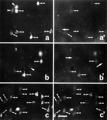
将TMR注入脑干运动前神经元所在区域后,TMR逆标神经元见于额叶1区和/或2区的V层,大脑皮质的其他部位几乎未见TMR逆标神经元,但将TMR注入脑干的不同部位后,TMR逆标神经元在运动皮质内的分布有明显的区别。将TMR注入臂旁外侧核后,TMR逆标神经元主要见于额叶2区吻端的V层(图a);将TMR注入三叉上核后,TMR逆标神经元主要见于额叶1区的V层(图b);将TMR注入延髓网状结构外侧部后,在额叶1区和2区的V层内均能见到TMR逆标神经元(图c)。PAG样阳性神经元见于大脑运动皮质Ⅱ~Ⅵ层的锥体细胞,PAG样免疫荧光染色阳性产物多呈颗粒状,仅见于阳性神经元的胞体,少数PAG样阳性细胞的染色较弱,其他类型的大脑运动皮质神经元几乎均为PAG样阴性(图a',b',c')。将TMR注入上述区域后,额叶1区和2区内的TMR逆标神经元几乎均呈PAG样阳性,但少数PAG样阳性锥体细胞的染色较弱(图a,a';b,b';c,c')。
, 百拇医药
图a 将四甲基罗达明(TMR)注入臂旁外侧核后,TMR逆标锥体细胞在吻端额叶2区V层内的分布 标尺示120μm(下同)
图b 将TMR注入三叉上核后,TMR逆标锥体细胞在额叶1区V层内的分布
图c 将TMR注入延髓网状结构外侧部后,TMR逆标锥体细胞在额叶1区V层内的分布
图a',b',c' PAG样阳性锥体细胞在额叶2区(a')和1区(b',c')V层内的分布
图a和a',b和b'及c和c'分别为同一视野在不同激发波长和发射波长时的照片,其中的双三角示TMR逆标并呈PAG样阳性的双标锥体细胞,单三角示TMR单标的锥体细胞,箭头示单纯PAG样阳性的锥体细胞
Fig. a Distribution of the tetramethyl rhodamine(TMR) retrogradely labeled pyramidal cells in the layer V of area 2 of the rostral frontal cortex after injecting TMR into the lateral parabrachial nucleus.
, http://www.100md.com
Fig. b Distribution of the TMR retrogradely labeled pyramidal cells in the layer V of area 1 of frontal cortex after injecting TMR into the supratrigeminal nucleus.
Fig. c Distribution of the TMR retrogradely labeled pyramidal cells in the layer V of area 1 of frontal cortex after injecting TMR into the lateral region of the medullary reticular formation.
Fig. a',b',c' Distributions of phosphate-activated glutaminase (PAG)-like immunoreactive pyramidal cells in the layer V of area 2 (a') and area 1(b',c')of frontal cortex.
, 百拇医药
Fig. a and a',b and b',c and c' are photographes taken from the same field under different wave lengths of exicitation and emission. In Figs. a and a',b and b',c and c', double-triangles indicate double-labeled pyramidal cells which were labeled with TMR and also exhibited PAG-like immunoreactivities, single-triangles indicate TMB single-labeled pyramidal cells, arrows indicate PAG-LI pyramidal cells. Bar in Fig. c' is 120 μm and also for Fig. a, a',b,b'/,and c.
, 百拇医药 本研究的结果说明,由大脑运动皮质V层锥体细胞发出的下行投射纤维所组成的皮质脑干束几乎均含兴奋性神经递质谷氨酸,皮质脑干束主要终止于脑干面口部运动前神经元所在的区域,但大脑运动皮质向不同脑干面口部运动前神经元所在区域投射的起源有差异,如额叶2区、额叶1区及额叶1区和2区V层锥体细胞的下行谷氨酸能投射主要分别终止于脑干的臂旁外侧核、三叉上核及延髓网状结构外侧部,这些部位是脑干运动前神经元所在的主要区域[5]。
本研究观察到大脑运动皮质发出下行投射的神经元均含谷氨酸,这对大脑运动皮质的运动指令下传是非常重要的。大脑运动皮质的下行投射并非直接终止于脑干运动核,而是先终止于脑干的运动前神经元,再经运动前神经元调控脑干面口运动核内的运动神经元。本研究的结果提示,脑干面口部运动前神经元主要接受大脑运动皮质兴奋性传入的调控。在面口部一系列精细运动中,需要面口部不同肌群的协同和协调,脑干面口部运动神经元在完成这些协同和协调的过程中,仅接受兴奋性传入显然是不足的,必然要有抑制性运动前神经元的参与。我们以往的研究已证实向脑干面口部运动核投射的运动前神经元含5-羟色胺、P物质和脑啡肽及抑制性神经递质γ-氨基丁酸和甘氨酸,这是完成上述运动调控的物质基础。
, 百拇医药
收稿 1997-08 修回 1998-06
参考文献
[1]Nozaki S, Iriki A, Nakamura Y. Trigeminal premotor neurons in the bulbar parvicellular reticular formation participating in induction of rhythmical activity of trigeminal motoneurons by repetitive stimulation of the cerebral cortex in the guinea pig. J Neurophysiol, 1993,69(3):595
[2]Yasui Y, Itoh K, Mitani A, et al. Cerebral cortical projections to the reticular regions around the trigeminal motor nucleus in the cat. J Comp Neurol, 1985;241(2):348
, http://www.100md.com
[3]Bradford HE, Ward HK, Thomas AJ. Glutamine——A major substrate for nerve endings. J Neurochem, 1978;30(4):1453
[4]Kaneko T, Mizuno N. Immunohistochemical study of glutaminase-containing neurons in the cerebral cortex and thalamus of the rat. J Comp Neurol, 1988;267(4);590
[5]李云庆,王智明,施际武.大鼠大脑运动皮质与脑干面口部运动前神经元的突触联系.解剖学报,1999,30(1)(待发表)
[6]Zilles K, Wree A. Cortex: areal and laminar structure. In: Paxinox G, (ed). The Rat Nervous System. ed2. San Diego: Academic Press, 1995:649-685
, http://www.100md.com
GLUTAMATERGIC PROJECTIONS FROM THE CEREBRAL
MOTOR CORTEX TO THE PREMOTOR NEURON POOL OF
THE BRAINSTEM IN THE RAT
MOTOR CORTEX TO THE PREMOTOR NEURON POOL OF
THE BRAINSTEM IN THE RAT
Li Yunqing△,Wang Zhiming, Shi Jiwu
(Department of Anatomy, K. K. Leung Brain Research Centre,The Fourth Military Medical University, Xi'an)
, 百拇医药
A retrograde tracing combined with immunoflurescent histochemical staining double-labeling technique was used in the present study to investagate the glutamatergic projections from the cerebral motor cortex to the premotor neuron pool of the brainstem. After injecting tetramethyl rhodamine (TMR) into the lateral parabrachial nucleus, supratrigmeinal nucleus, or lateral region of the medullary reticular formation, TMR retrogradely labeled pyramidal cells were found in the layer V of the area 2, area 1, or area 1 and area 2 of the frontal cortex, respectively. Phosphate-activated glutaminase-like immunoreactive (PAG-LI) pyramidal cells were observed through out layers Ⅱ~Ⅵ of the cerebral motor cortex. Almost all of the TMR retrogradely labeled pyramidal cells also showed PAG-like immunoreactivities. The present results indicate that: (1) corticobulbar tract mainly terminates in the premotor neuron pool of the brainstem; (2) different subregions of the premotor neuron pool of the brainstem receive afferent projections originated from different subregions of the cerebral motor cortex; (3) glutamate is the major neurotransmitter of the corticobulbar tract.
KEY WORDS Cerebral motor cortex; Layer V; Corticobulbar tract; Premotor neuron; Glutamate; Rat
△Department of Anatomy, K. K. Leung Brain Research Centre. The Fourth Military Medical University, Xi'an 710032, China
*国家杰出青年科学基金资助课题(No.39625011), 百拇医药
单位:第四军医大学解剖学教研室、梁琚脑研究中心,西安 710032
关键词:运动皮质;V层;皮质脑干束;运动前神经元;谷氨酸;大鼠
解剖学报/980423 摘 要 为观察大鼠大脑运动皮质向脑干运动前神经元所在区域的谷氨酸能投射。用逆行追踪和免疫荧光组织化学染色相结合的双标技术,将四甲基罗达明(TMR)分别注入臂旁外侧核、三叉上核或延髓网状结构外侧部后,TMR逆标神经元分别见于额叶2区、额叶1区或额叶1区和2区的V层;磷酸激活的谷氨酰胺酶(PAG)样阳性神经元见于大脑运动皮质Ⅱ~Ⅵ层的锥体细胞;TMR逆标神经元几乎均呈PAG样阳性。结果提示,大脑运动皮质发出的皮质脑干束主要终止于脑干运动前神经元所在的区域,运动皮质向不同的脑干运动前神经元所在区域发出投射的起源也有差异,谷氨酸是此通路的主要神经递质。
, 百拇医药
大脑运动皮质V层锥体细胞的传出纤维所组成的皮质脑干束主要终止于脑干的运动前神经元,经过运动前神经元调控脑干面口部运动核内运动神经元的活动,从而达到对面口部不同肌群间的协同和协调,完成一系列复杂的精细运动[1,2]。谷氨酸是大脑皮质锥体细胞的主要兴奋性神经递质,磷酸激活的谷氨酰胺酶(phosphate-activated glutaminase, PAG)是合成谷氨酸的关键酶,所以PAG可以用作中枢内谷氨酸样阳性神经元的特异性标记物[3,4],我们的研究观察到谷氨酸样阳性终末与向脑干面口部运动核投射的脑干运动前神经元形成非对称性突触联系[5],但这些谷氨酸样阳性终末的来源和大脑运动皮质的谷氨酸能下行投射尚缺乏系统观察。大鼠的运动皮质是额叶1区和2区[6]。本研究用四甲基罗达明(tetramethyl rhodamin, TMR)逆行追踪与PAG样免疫荧光组织化学染色相结合的双标技术,对大鼠大脑运动皮质向脑干运动前神经元所在区域的谷氨酸能下行投射进行了观察。
, http://www.100md.com
材料和方法
SD雄性大鼠30只,体重200~250g。戊巴比妥钠麻醉(40 mg/kg,i.p.)后,将0.1 μl的3%TMR(molecular probe)用尖端粘微玻管的微量注射器经压力分别注入臂旁外侧核(10只)、三叉上核(10只)和延髓网状结构外侧部(10只),注射时间20 min,注毕留针20 min。动物存活72h后用过量戊巴比妥钠深麻,开胸,插管至升主动脉,按Kaneko等[4]的方法,先以100 ml生理盐水冲洗血液,再用500 ml含0.2%福尔马林和饱和苦味酸的0.1 mol/L磷酸盐缓冲液(PB,pH 7.0)灌注固定1h。灌注完毕立即取脑,用含2%福尔马林和饱和苦味酸的新鲜固定液后固定24h(4℃),再移入30%蔗糖中至沉底(4℃)。冠状冻切前脑和脑干,片厚20μm,切片隔2张取1张,分3组收集于0.01 mol/L PBS(pH7.4)中。将第1组切片裱于载片上,干燥后用荧光显微镜观察。选注射区位于臂旁外侧核(6只)、三叉上核(6只)和延髓网状结构外侧部(6只)内的18只动物的切片,行PAG免疫荧光组织化学染色,具体步骤为:(1) 每只动物的第2组切片入小鼠IgM抗PAG血清(1∶1 000,日本京都大学金子武嗣先生惠赠)[4],室温孵育24h;(2) Biotin标记的驴抗小鼠IgM血清(1∶200,Jackson),室温孵育3h;(3)Avidin结合的FITC(1∶1 000,Vector),室温孵育2h。裱片、避光干燥、甘油和PBS(1∶1)混合液封片。在荧光显微镜下观察、计数并摄片。选择激发波长为540~552 nm和发射波长为≥580 nm的滤色片观察桔红色的TMR标记神经元,激发波长为450~490 nm和发射波长为515~565 nm的滤色片观察FITC标记的绿色PAG样免疫荧光染色阳性神经元。另一组切片用于Nissl染色,以便对照观察和定位。
, http://www.100md.com
结果和讨论
将TMR注入脑干运动前神经元所在区域后,TMR逆标神经元见于额叶1区和/或2区的V层,大脑皮质的其他部位几乎未见TMR逆标神经元,但将TMR注入脑干的不同部位后,TMR逆标神经元在运动皮质内的分布有明显的区别。将TMR注入臂旁外侧核后,TMR逆标神经元主要见于额叶2区吻端的V层(图a);将TMR注入三叉上核后,TMR逆标神经元主要见于额叶1区的V层(图b);将TMR注入延髓网状结构外侧部后,在额叶1区和2区的V层内均能见到TMR逆标神经元(图c)。PAG样阳性神经元见于大脑运动皮质Ⅱ~Ⅵ层的锥体细胞,PAG样免疫荧光染色阳性产物多呈颗粒状,仅见于阳性神经元的胞体,少数PAG样阳性细胞的染色较弱,其他类型的大脑运动皮质神经元几乎均为PAG样阴性(图a',b',c')。将TMR注入上述区域后,额叶1区和2区内的TMR逆标神经元几乎均呈PAG样阳性,但少数PAG样阳性锥体细胞的染色较弱(图a,a';b,b';c,c')。

, 百拇医药
图a 将四甲基罗达明(TMR)注入臂旁外侧核后,TMR逆标锥体细胞在吻端额叶2区V层内的分布 标尺示120μm(下同)
图b 将TMR注入三叉上核后,TMR逆标锥体细胞在额叶1区V层内的分布
图c 将TMR注入延髓网状结构外侧部后,TMR逆标锥体细胞在额叶1区V层内的分布
图a',b',c' PAG样阳性锥体细胞在额叶2区(a')和1区(b',c')V层内的分布
图a和a',b和b'及c和c'分别为同一视野在不同激发波长和发射波长时的照片,其中的双三角示TMR逆标并呈PAG样阳性的双标锥体细胞,单三角示TMR单标的锥体细胞,箭头示单纯PAG样阳性的锥体细胞
Fig. a Distribution of the tetramethyl rhodamine(TMR) retrogradely labeled pyramidal cells in the layer V of area 2 of the rostral frontal cortex after injecting TMR into the lateral parabrachial nucleus.
, http://www.100md.com
Fig. b Distribution of the TMR retrogradely labeled pyramidal cells in the layer V of area 1 of frontal cortex after injecting TMR into the supratrigeminal nucleus.
Fig. c Distribution of the TMR retrogradely labeled pyramidal cells in the layer V of area 1 of frontal cortex after injecting TMR into the lateral region of the medullary reticular formation.
Fig. a',b',c' Distributions of phosphate-activated glutaminase (PAG)-like immunoreactive pyramidal cells in the layer V of area 2 (a') and area 1(b',c')of frontal cortex.
, 百拇医药
Fig. a and a',b and b',c and c' are photographes taken from the same field under different wave lengths of exicitation and emission. In Figs. a and a',b and b',c and c', double-triangles indicate double-labeled pyramidal cells which were labeled with TMR and also exhibited PAG-like immunoreactivities, single-triangles indicate TMB single-labeled pyramidal cells, arrows indicate PAG-LI pyramidal cells. Bar in Fig. c' is 120 μm and also for Fig. a, a',b,b'/,and c.
, 百拇医药 本研究的结果说明,由大脑运动皮质V层锥体细胞发出的下行投射纤维所组成的皮质脑干束几乎均含兴奋性神经递质谷氨酸,皮质脑干束主要终止于脑干面口部运动前神经元所在的区域,但大脑运动皮质向不同脑干面口部运动前神经元所在区域投射的起源有差异,如额叶2区、额叶1区及额叶1区和2区V层锥体细胞的下行谷氨酸能投射主要分别终止于脑干的臂旁外侧核、三叉上核及延髓网状结构外侧部,这些部位是脑干运动前神经元所在的主要区域[5]。
本研究观察到大脑运动皮质发出下行投射的神经元均含谷氨酸,这对大脑运动皮质的运动指令下传是非常重要的。大脑运动皮质的下行投射并非直接终止于脑干运动核,而是先终止于脑干的运动前神经元,再经运动前神经元调控脑干面口运动核内的运动神经元。本研究的结果提示,脑干面口部运动前神经元主要接受大脑运动皮质兴奋性传入的调控。在面口部一系列精细运动中,需要面口部不同肌群的协同和协调,脑干面口部运动神经元在完成这些协同和协调的过程中,仅接受兴奋性传入显然是不足的,必然要有抑制性运动前神经元的参与。我们以往的研究已证实向脑干面口部运动核投射的运动前神经元含5-羟色胺、P物质和脑啡肽及抑制性神经递质γ-氨基丁酸和甘氨酸,这是完成上述运动调控的物质基础。
, 百拇医药
收稿 1997-08 修回 1998-06
参考文献
[1]Nozaki S, Iriki A, Nakamura Y. Trigeminal premotor neurons in the bulbar parvicellular reticular formation participating in induction of rhythmical activity of trigeminal motoneurons by repetitive stimulation of the cerebral cortex in the guinea pig. J Neurophysiol, 1993,69(3):595
[2]Yasui Y, Itoh K, Mitani A, et al. Cerebral cortical projections to the reticular regions around the trigeminal motor nucleus in the cat. J Comp Neurol, 1985;241(2):348
, http://www.100md.com
[3]Bradford HE, Ward HK, Thomas AJ. Glutamine——A major substrate for nerve endings. J Neurochem, 1978;30(4):1453
[4]Kaneko T, Mizuno N. Immunohistochemical study of glutaminase-containing neurons in the cerebral cortex and thalamus of the rat. J Comp Neurol, 1988;267(4);590
[5]李云庆,王智明,施际武.大鼠大脑运动皮质与脑干面口部运动前神经元的突触联系.解剖学报,1999,30(1)(待发表)
[6]Zilles K, Wree A. Cortex: areal and laminar structure. In: Paxinox G, (ed). The Rat Nervous System. ed2. San Diego: Academic Press, 1995:649-685
, http://www.100md.com
GLUTAMATERGIC PROJECTIONS FROM THE CEREBRAL
MOTOR CORTEX TO THE PREMOTOR NEURON POOL OF
THE BRAINSTEM IN THE RAT
MOTOR CORTEX TO THE PREMOTOR NEURON POOL OF
THE BRAINSTEM IN THE RAT
Li Yunqing△,Wang Zhiming, Shi Jiwu
(Department of Anatomy, K. K. Leung Brain Research Centre,The Fourth Military Medical University, Xi'an)
, 百拇医药
A retrograde tracing combined with immunoflurescent histochemical staining double-labeling technique was used in the present study to investagate the glutamatergic projections from the cerebral motor cortex to the premotor neuron pool of the brainstem. After injecting tetramethyl rhodamine (TMR) into the lateral parabrachial nucleus, supratrigmeinal nucleus, or lateral region of the medullary reticular formation, TMR retrogradely labeled pyramidal cells were found in the layer V of the area 2, area 1, or area 1 and area 2 of the frontal cortex, respectively. Phosphate-activated glutaminase-like immunoreactive (PAG-LI) pyramidal cells were observed through out layers Ⅱ~Ⅵ of the cerebral motor cortex. Almost all of the TMR retrogradely labeled pyramidal cells also showed PAG-like immunoreactivities. The present results indicate that: (1) corticobulbar tract mainly terminates in the premotor neuron pool of the brainstem; (2) different subregions of the premotor neuron pool of the brainstem receive afferent projections originated from different subregions of the cerebral motor cortex; (3) glutamate is the major neurotransmitter of the corticobulbar tract.
KEY WORDS Cerebral motor cortex; Layer V; Corticobulbar tract; Premotor neuron; Glutamate; Rat
△Department of Anatomy, K. K. Leung Brain Research Centre. The Fourth Military Medical University, Xi'an 710032, China
*国家杰出青年科学基金资助课题(No.39625011), 百拇医药