心理应激对正常人外周血T淋巴细胞体外活化表面分子表达的作用
作者:黄柏炎 孙荭 曾洁铭 程云华 王曦 曾耀英
单位:黄柏炎(暨南大学生物工程学系, 广东 广州 510632);孙荭 曾洁铭(暨南大学组织移植与免疫中心);程云华(暨南大学医学院第一附院);王曦 曾耀英(暨南大学组织移植与免疫中心)
关键词:心理学,医学;T淋巴细胞;抗原,CD69
中国病理生理杂志000311
[摘 要] 目的:探讨心理应激增加对感染的易感性的细胞和分子免疫学机理。方法:采用双荧光染色流式细胞分析法对20名健康大学生志愿者(男女各半)在应激前后进行外周血淋巴细胞免疫表型及T细胞体外丝裂原刺激早期活化抗原表达分析。连续一周期末考试被设定为心理应激。结果:免疫表型分析显示,应激前后CD2、CD3、CD4、CD8、CD19、CD20、CD16、CD56等淋巴细胞的表面分子阳性的细胞的百分比的差异无统计学显著性;与应激前的结果相比,应激后的T细胞在体外培养条件下多克隆刺激剂活化后CD69的表达明显降低,植物血凝素(phytohemagglutinin,PHA)组CD69+CD3+/CD3+的百分率由应激前的28.1±4.1降低到应激后的17.6±3.8,佛波醇酯(phorbol 12,13-dibutyrate,PDB)组CD69+CD3+/CD3+的百分率由应激前的80.7±6.8降至应激后的65.8±7.9,而在没有刺激剂作用的条件下,T细胞CD69表达率应激前后的差别无显著。结论:应激对免疫系统的影响并不在于改变外周血淋巴细胞各亚群的比例的层面上;心理应激能降低健康人T细胞体外活化的反应性,这可能与心理应激个体对感染的易感性增加有关。
, 百拇医药
[中图分类号] R392.9 [文献标识码] A
[文章编号] 1000-4718(2000)03-0229-04
Effects of psychological stress on in vitro expression of activated surface molecules on T cells of peripheral blood from healthy persons
HUANG Bo-yan
(Department of Biotechnology, Jinan University, Guangzhou 510632, China)
SUN Hong, ZENG Jie-ming, WANG Xi, ZENG Yao-ying
, http://www.100md.com
(Institute for Tissue Transplantation & Immunology, Jinan University)
CHENG Yun-hua
(First Affiliated Hospital of Jinan University Medical College)
[Abstract] AIM: To study cellular and molecular mechanism involved in increasing susceptibility of infection in psychological stress persons. METHODS: Comparative studies were performed with double staining and flow cytometry analysis on immunophenotyping and in vitro expression of early activating surface molecule CD69 in response to mitogens on T cells from peripheral blood of 20 healthy college student volunteers before and after psychological stress. A series of term final examinations was defined as psychological stress. RESULTS: Immunophenotyping analysis showed no statistically significant difference in the percentage of CD2, CD3, CD4, CD8, CD19, CD20, CD16 and CD56 positive lymphocyte populations before and after psychological stress. There was a statistically significant decrease in the in vitro expression of CD69 in response to polyclonal stimulators on the T cells from persons after psychological stress than those before psychological stress. The percentage of CD69 expression (CD69+CD3+/CD3+%) in response to PHA and PDB in the whole blood culture for 72 hours decreased respectively from 28.1±4.1 and 80.7±6.8 on the T cells obtained before psychological stress to 17.6±3.8 and 65.8±7.9 on those obtained after psychological stress, while there was no statistically significant difference between the CD69 expression rates without stimulators on the T cells obtained before and after psychological stress. CONCLUSIONS: The effects of psychological stress to immune system is not on the level of changing proportions of the sub-populations within peripheral blood lymphocytes. Psychological stress can decrease the activating response of T cells in healthy persons, which may be responsible for the increase of susceptibility to infection in the psychological stress persons.
, http://www.100md.com
[MeSH] Psychological, medical; T-lymphocytes; Antigens, CD69
有证据表明:脑、内分泌系统和免疫系统共享同一套细胞因子、肽激素、神经递质及相应受体,因而这些分子就成为神经内分泌和免疫系统进行双向信息交流的化学语言[1,2]。虽然人们早已观察到心理应激(psychological stress)能增加个体对微生物感染的易感性[3],但其细胞和分子机制尚未明了。本研究旨在探讨心理应激增加人体对感染的易感性的细胞和分子免疫学机制。
材 料 和 方 法
一、研究对象
20名健康大学生志愿者参加此项研究,其中男、女学生各10名,年龄为17~22岁。连续一周的期末考试被设定为心理应激的刺激, 被调查者兼担心所要面临的考试难度较大, 有不及格的危险。考试开始前1周所抽取的静脉血为心理应激前样品,经历1周考试后所抽取的静脉血为心理应激后样品。为排除不同时间免疫表型及体外活化表面分子表达的差别,我们在学生准备考试前3周已连续2次对他们进行试验测定, 以排除其他心理应激的可能性。其中受检女学生兼排除月经期,以排除激素水平变化的影响。
, 百拇医药
二、单克隆抗体及试剂
本研究所使用的CD69等单克隆抗体购自法国IMMUNOTECH公司或美国Becton Dickinson 公司,PHA、PDB等多克隆刺激剂购自美国Sigma公司,RPMI-1640、胎牛血清(fetal bovine serum, FBS)等细胞培养试剂购自美国Gibco公司。
三、实验方法
荧光染色流式细胞分析法用于外周血淋巴细胞免疫表型及T细胞体外丝裂原刺激早期活化抗原表达分析[4,5]。间接荧光标记法被用于淋巴细胞免疫表型分析,程序步骤如下: 30μL肝素抗凝静脉血与10μL第一单抗(鼠抗人)混合后室温孵育30 min,然后用红细胞裂解液裂解红细胞,离心获取有核细胞,PBS洗涤后加入荧光标记的羊抗鼠单抗(第二单抗),混合后室温孵育30 min,PBS洗涤1次,用1%多聚甲醛固定10 min后,用流式细胞仪进行分析。间接-直接双荧光标记法被用于T细胞活化CD69表达的检测,程序步骤与间接标记法相似,只是在用多聚甲醛固定前插入以下步骤:加入荧光标记的单抗后室温孵育30 min并用PBS洗涤1次。T细胞活化采取体外全血培养多克隆活化剂刺激法,抗凝全血用2倍完全RPMI-1640培养液稀释,在CO2孵箱培养72 h。全部数据用FACStarplus流式细胞仪和LYSYS Ⅱ软件进行单荧光或双荧光参数获取和分析。
, http://www.100md.com
结 果
一、非考试同期自身对照及心理应激前后免疫表型分析
考试应激前后及非考试同期自身对照CD2、CD3、CD4、CD8、CD19、CD20、CD16、CD56等淋巴细胞的表面分子阳性的细胞的百分比的差异无显著差别(见表1)。
二、非考试同期自身对照及心理应激前后T细胞体外活化分析
与应激前的结果相比,应激后的T细胞在体外培养条件下多克隆刺激剂活化后CD69的表达明显降低。而非考试同期自身对照CD69表达百分比的差异无显著。体外全血培养72 h,PHA组CD69+CD3+/CD3+的百分率由应激前的28.1±4.1降低到应激后的17.6±3.8,PDB组CD69+CD3+/CD3+的百分率由应激前的80.7±6.8降至应激后的65.8±7.9,在没有刺激剂作用的条件下,T细胞CD69表达率应激前后的差别无显著(见表2)。图1显示心理应激前后T细胞体外活化后用流式细胞仪进行分析时,各种情况下的实际记录。
, 百拇医药
表1 非考试同期自身对照及心理应激前后淋巴细胞免疫表型分析
Tab 1 Immunophenotyping of lymphocytes before and after psychological stress in normal persons ( ±s,n=20)
T cells
B cells
NK cells
CD2
CD3
CD4
, 百拇医药 CD8
CD19
CD23
CD16
CD56
Control
Stress
First
week
74.9±8.3
66.1±6.6
42.7±6.7
27.7±6.9
, 百拇医药
6.5±2.7
2.3±1.4
13.7±5.8
14.5±5.2
Second
week
73.8±7.9
65.2±7.0
48.0±7.2
25.7±7.1
7.2±2.1
3.1±1.8
, http://www.100md.com
14.2±4.6
15.6±6.4
Before
Pys.str
75.0±8.0△
64.1±7.5△
44.6±6.8△
26.8±6.5△
5.6±1.9△
2.5±1.7△
, http://www.100md.com
12.8±4.8△
13.8±4.8△
After
Pys.str
74.6±8.8*
67.0±6.1*
44.6±6.0*
25.8±7.2*
7.0±1.8*
1.8±2.3*
, 百拇医药
14.8±6.2*
12.7±5.4*
△P>0.05, vs control group; *P>0.05, vs before psychological stress; Pys.str=psychological stress表2 非考试同期自身对照及心理应激
前后T细胞体外活化CD69表达的作用
Tab 1 Immunophenotyping of lymphocytes before and after
psychological stress in normal persons (±s,n=20)
, 百拇医药
Stimulators
None
PHA
PDB
Control
Stress
First
week
1.5±0.8
26.7±3.9△
81.0±7.1*
Second week
, 百拇医药
1.4±0.9
27.6±3.5△
82.3±8.3*
Before
Pys.str
1.6±1.1
28.1±4.1△
80.7±6.8*
After
Pys.str
1.3±0.7
, 百拇医药
17.6±3.8△△
65.8±7.9**
△P>0.05, vs each other; *P>0.05, vs each other; △△P<0.01, vs PHA-stimulating group before psychological stress; **P<0.01,vs PDB-stimulating group before psychological stress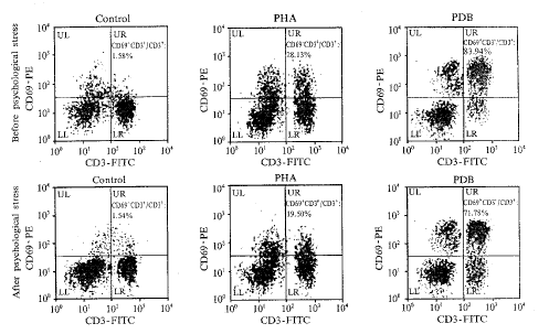
Fig 1 Effect of psychological stress on expression of CD69 by T cells during in vitro activation
, http://www.100md.com 图1 心理应激对T细胞体外活化CD69表达的作用
讨 论
人外周血淋巴细胞为非均一的细胞群,它包括T细胞、B细胞和NK细胞等细胞群,而T细胞又可分为T辅助细胞(T helper, Th)和细胞毒T细胞(cytolytic T lymphocytes, CTL)等若干亚群。在正常情况下,各群细胞按一定的比例形成某种正常布局,而在某些疾病状态下这种正常布局会发生偏移。每群淋巴细胞都有特征性的细胞表面标记分子,因而免疫表型分析的结果可揭示各群细胞的相对比例。免疫表型分析结果显示:应激前后总T细胞、Th、CTL、B细胞和NK细胞的比例差异无显著,这说明应激对免疫系统的影响不在于改变外周血淋巴细胞各亚群相对比例的层面上。T细胞在整个免疫应答中起着关键性作用,而T细胞的活化效能是T细胞功能的反映。在某些疾病状态下,T细胞的活化效能会发生改变。T细胞的活化效能可以通过在体外条件下对多克隆刺激剂的反应来检测。CD69是T细胞活化后表达的细胞表面分子[6~8],用荧光抗体对CD3和CD69进行双色标记后,再用流式细胞仪进行分析可以准确地检测活化T细胞的相对数。我们的结果显示,与应激前的结果相比,应激后的T细胞在体外培养条件下,经多克隆刺激剂活化后,CD69的表达明显降低,而在没有刺激剂作用的条件下,T细胞CD69的表达率在应激前后的差别无显著。我们的结果提示,心理应激能降低健康人T细胞的活化反应潜能,这可能与心理应激个体感染的易感性增加有关。
, http://www.100md.com
[基金项目] 国家自然科学基金资助(39670690);广东省自然科学基金资助(960196)
[参 考 文 献]
[1] Blalock JE. The syntax of immune-neuroendocrine communication [J]. Immunol Today, 1994, 15(11): 504~511.
[2] Sternberg EM. Neural-immune interactions in health and disease [J]. J Clin Invest, 1997, 100(11): 2641~2647.
[3] Brines R. Neuroendocine immunology today [J]. Immunol Today,1994,15(11): 503.
, http://www.100md.com
[4] 蔡小嫦,孙荭,曾耀英,等. 白癜风患者外周血T淋巴细胞异常活化 [J]. 中国病理生理杂志,1994,10(5):487~492.
[5] 孙荭,曾耀英,葛琪,等. 口服人参水煎剂对正常人淋巴细胞免疫表型及体外活化IL-2R表达的作用 [J]. 暨南大学学报(医学版),1996,17s:88~90.
[6] Mehta BA, Maino VC. Simultaneous detection of DNA synthesis and cytokine production in staphylococcal enterotoxin B activated CD4+ T lymphocytes by flow cytometry [J]. J Immunol Methods, 1997, 208: 49~59.
[7] Craston R, Koh M, Dermott AM, et al. Temporal dynamics of CD69 expression on lymphoid cells [J]. J Immunol Methods, 1997, 209: 37~45.
[8] Testi R, Ambrosio D, Maria R, et al. The CD69 receptor: a multipurpose cell surface trigger for hematopoitic cells [J]. Immunol Today, 1994, 15(10): 479~483.
[收稿日期]1998-10-26 [修回日期]1999-12-28
, 百拇医药
单位:黄柏炎(暨南大学生物工程学系, 广东 广州 510632);孙荭 曾洁铭(暨南大学组织移植与免疫中心);程云华(暨南大学医学院第一附院);王曦 曾耀英(暨南大学组织移植与免疫中心)
关键词:心理学,医学;T淋巴细胞;抗原,CD69
中国病理生理杂志000311
[摘 要] 目的:探讨心理应激增加对感染的易感性的细胞和分子免疫学机理。方法:采用双荧光染色流式细胞分析法对20名健康大学生志愿者(男女各半)在应激前后进行外周血淋巴细胞免疫表型及T细胞体外丝裂原刺激早期活化抗原表达分析。连续一周期末考试被设定为心理应激。结果:免疫表型分析显示,应激前后CD2、CD3、CD4、CD8、CD19、CD20、CD16、CD56等淋巴细胞的表面分子阳性的细胞的百分比的差异无统计学显著性;与应激前的结果相比,应激后的T细胞在体外培养条件下多克隆刺激剂活化后CD69的表达明显降低,植物血凝素(phytohemagglutinin,PHA)组CD69+CD3+/CD3+的百分率由应激前的28.1±4.1降低到应激后的17.6±3.8,佛波醇酯(phorbol 12,13-dibutyrate,PDB)组CD69+CD3+/CD3+的百分率由应激前的80.7±6.8降至应激后的65.8±7.9,而在没有刺激剂作用的条件下,T细胞CD69表达率应激前后的差别无显著。结论:应激对免疫系统的影响并不在于改变外周血淋巴细胞各亚群的比例的层面上;心理应激能降低健康人T细胞体外活化的反应性,这可能与心理应激个体对感染的易感性增加有关。
, 百拇医药
[中图分类号] R392.9 [文献标识码] A
[文章编号] 1000-4718(2000)03-0229-04
Effects of psychological stress on in vitro expression of activated surface molecules on T cells of peripheral blood from healthy persons
HUANG Bo-yan
(Department of Biotechnology, Jinan University, Guangzhou 510632, China)
SUN Hong, ZENG Jie-ming, WANG Xi, ZENG Yao-ying
, http://www.100md.com
(Institute for Tissue Transplantation & Immunology, Jinan University)
CHENG Yun-hua
(First Affiliated Hospital of Jinan University Medical College)
[Abstract] AIM: To study cellular and molecular mechanism involved in increasing susceptibility of infection in psychological stress persons. METHODS: Comparative studies were performed with double staining and flow cytometry analysis on immunophenotyping and in vitro expression of early activating surface molecule CD69 in response to mitogens on T cells from peripheral blood of 20 healthy college student volunteers before and after psychological stress. A series of term final examinations was defined as psychological stress. RESULTS: Immunophenotyping analysis showed no statistically significant difference in the percentage of CD2, CD3, CD4, CD8, CD19, CD20, CD16 and CD56 positive lymphocyte populations before and after psychological stress. There was a statistically significant decrease in the in vitro expression of CD69 in response to polyclonal stimulators on the T cells from persons after psychological stress than those before psychological stress. The percentage of CD69 expression (CD69+CD3+/CD3+%) in response to PHA and PDB in the whole blood culture for 72 hours decreased respectively from 28.1±4.1 and 80.7±6.8 on the T cells obtained before psychological stress to 17.6±3.8 and 65.8±7.9 on those obtained after psychological stress, while there was no statistically significant difference between the CD69 expression rates without stimulators on the T cells obtained before and after psychological stress. CONCLUSIONS: The effects of psychological stress to immune system is not on the level of changing proportions of the sub-populations within peripheral blood lymphocytes. Psychological stress can decrease the activating response of T cells in healthy persons, which may be responsible for the increase of susceptibility to infection in the psychological stress persons.
, http://www.100md.com
[MeSH] Psychological, medical; T-lymphocytes; Antigens, CD69
有证据表明:脑、内分泌系统和免疫系统共享同一套细胞因子、肽激素、神经递质及相应受体,因而这些分子就成为神经内分泌和免疫系统进行双向信息交流的化学语言[1,2]。虽然人们早已观察到心理应激(psychological stress)能增加个体对微生物感染的易感性[3],但其细胞和分子机制尚未明了。本研究旨在探讨心理应激增加人体对感染的易感性的细胞和分子免疫学机制。
材 料 和 方 法
一、研究对象
20名健康大学生志愿者参加此项研究,其中男、女学生各10名,年龄为17~22岁。连续一周的期末考试被设定为心理应激的刺激, 被调查者兼担心所要面临的考试难度较大, 有不及格的危险。考试开始前1周所抽取的静脉血为心理应激前样品,经历1周考试后所抽取的静脉血为心理应激后样品。为排除不同时间免疫表型及体外活化表面分子表达的差别,我们在学生准备考试前3周已连续2次对他们进行试验测定, 以排除其他心理应激的可能性。其中受检女学生兼排除月经期,以排除激素水平变化的影响。
, 百拇医药
二、单克隆抗体及试剂
本研究所使用的CD69等单克隆抗体购自法国IMMUNOTECH公司或美国Becton Dickinson 公司,PHA、PDB等多克隆刺激剂购自美国Sigma公司,RPMI-1640、胎牛血清(fetal bovine serum, FBS)等细胞培养试剂购自美国Gibco公司。
三、实验方法
荧光染色流式细胞分析法用于外周血淋巴细胞免疫表型及T细胞体外丝裂原刺激早期活化抗原表达分析[4,5]。间接荧光标记法被用于淋巴细胞免疫表型分析,程序步骤如下: 30μL肝素抗凝静脉血与10μL第一单抗(鼠抗人)混合后室温孵育30 min,然后用红细胞裂解液裂解红细胞,离心获取有核细胞,PBS洗涤后加入荧光标记的羊抗鼠单抗(第二单抗),混合后室温孵育30 min,PBS洗涤1次,用1%多聚甲醛固定10 min后,用流式细胞仪进行分析。间接-直接双荧光标记法被用于T细胞活化CD69表达的检测,程序步骤与间接标记法相似,只是在用多聚甲醛固定前插入以下步骤:加入荧光标记的单抗后室温孵育30 min并用PBS洗涤1次。T细胞活化采取体外全血培养多克隆活化剂刺激法,抗凝全血用2倍完全RPMI-1640培养液稀释,在CO2孵箱培养72 h。全部数据用FACStarplus流式细胞仪和LYSYS Ⅱ软件进行单荧光或双荧光参数获取和分析。
, http://www.100md.com
结 果
一、非考试同期自身对照及心理应激前后免疫表型分析
考试应激前后及非考试同期自身对照CD2、CD3、CD4、CD8、CD19、CD20、CD16、CD56等淋巴细胞的表面分子阳性的细胞的百分比的差异无显著差别(见表1)。
二、非考试同期自身对照及心理应激前后T细胞体外活化分析
与应激前的结果相比,应激后的T细胞在体外培养条件下多克隆刺激剂活化后CD69的表达明显降低。而非考试同期自身对照CD69表达百分比的差异无显著。体外全血培养72 h,PHA组CD69+CD3+/CD3+的百分率由应激前的28.1±4.1降低到应激后的17.6±3.8,PDB组CD69+CD3+/CD3+的百分率由应激前的80.7±6.8降至应激后的65.8±7.9,在没有刺激剂作用的条件下,T细胞CD69表达率应激前后的差别无显著(见表2)。图1显示心理应激前后T细胞体外活化后用流式细胞仪进行分析时,各种情况下的实际记录。
, 百拇医药
表1 非考试同期自身对照及心理应激前后淋巴细胞免疫表型分析
Tab 1 Immunophenotyping of lymphocytes before and after psychological stress in normal persons (
±s,n=20)T cells
B cells
NK cells
CD2
CD3
CD4
, 百拇医药 CD8
CD19
CD23
CD16
CD56
Control
Stress
First
week
74.9±8.3
66.1±6.6
42.7±6.7
27.7±6.9
, 百拇医药
6.5±2.7
2.3±1.4
13.7±5.8
14.5±5.2
Second
week
73.8±7.9
65.2±7.0
48.0±7.2
25.7±7.1
7.2±2.1
3.1±1.8
, http://www.100md.com
14.2±4.6
15.6±6.4
Before
Pys.str
75.0±8.0△
64.1±7.5△
44.6±6.8△
26.8±6.5△
5.6±1.9△
2.5±1.7△
, http://www.100md.com
12.8±4.8△
13.8±4.8△
After
Pys.str
74.6±8.8*
67.0±6.1*
44.6±6.0*
25.8±7.2*
7.0±1.8*
1.8±2.3*
, 百拇医药
14.8±6.2*
12.7±5.4*
△P>0.05, vs control group; *P>0.05, vs before psychological stress; Pys.str=psychological stress表2 非考试同期自身对照及心理应激
前后T细胞体外活化CD69表达的作用
Tab 1 Immunophenotyping of lymphocytes before and after
psychological stress in normal persons (
±s,n=20), 百拇医药
Stimulators
None
PHA
PDB
Control
Stress
First
week
1.5±0.8
26.7±3.9△
81.0±7.1*
Second week
, 百拇医药
1.4±0.9
27.6±3.5△
82.3±8.3*
Before
Pys.str
1.6±1.1
28.1±4.1△
80.7±6.8*
After
Pys.str
1.3±0.7
, 百拇医药
17.6±3.8△△
65.8±7.9**
△P>0.05, vs each other; *P>0.05, vs each other; △△P<0.01, vs PHA-stimulating group before psychological stress; **P<0.01,vs PDB-stimulating group before psychological stress
Fig 1 Effect of psychological stress on expression of CD69 by T cells during in vitro activation
, http://www.100md.com 图1 心理应激对T细胞体外活化CD69表达的作用
讨 论
人外周血淋巴细胞为非均一的细胞群,它包括T细胞、B细胞和NK细胞等细胞群,而T细胞又可分为T辅助细胞(T helper, Th)和细胞毒T细胞(cytolytic T lymphocytes, CTL)等若干亚群。在正常情况下,各群细胞按一定的比例形成某种正常布局,而在某些疾病状态下这种正常布局会发生偏移。每群淋巴细胞都有特征性的细胞表面标记分子,因而免疫表型分析的结果可揭示各群细胞的相对比例。免疫表型分析结果显示:应激前后总T细胞、Th、CTL、B细胞和NK细胞的比例差异无显著,这说明应激对免疫系统的影响不在于改变外周血淋巴细胞各亚群相对比例的层面上。T细胞在整个免疫应答中起着关键性作用,而T细胞的活化效能是T细胞功能的反映。在某些疾病状态下,T细胞的活化效能会发生改变。T细胞的活化效能可以通过在体外条件下对多克隆刺激剂的反应来检测。CD69是T细胞活化后表达的细胞表面分子[6~8],用荧光抗体对CD3和CD69进行双色标记后,再用流式细胞仪进行分析可以准确地检测活化T细胞的相对数。我们的结果显示,与应激前的结果相比,应激后的T细胞在体外培养条件下,经多克隆刺激剂活化后,CD69的表达明显降低,而在没有刺激剂作用的条件下,T细胞CD69的表达率在应激前后的差别无显著。我们的结果提示,心理应激能降低健康人T细胞的活化反应潜能,这可能与心理应激个体感染的易感性增加有关。
, http://www.100md.com
[基金项目] 国家自然科学基金资助(39670690);广东省自然科学基金资助(960196)
[参 考 文 献]
[1] Blalock JE. The syntax of immune-neuroendocrine communication [J]. Immunol Today, 1994, 15(11): 504~511.
[2] Sternberg EM. Neural-immune interactions in health and disease [J]. J Clin Invest, 1997, 100(11): 2641~2647.
[3] Brines R. Neuroendocine immunology today [J]. Immunol Today,1994,15(11): 503.
, http://www.100md.com
[4] 蔡小嫦,孙荭,曾耀英,等. 白癜风患者外周血T淋巴细胞异常活化 [J]. 中国病理生理杂志,1994,10(5):487~492.
[5] 孙荭,曾耀英,葛琪,等. 口服人参水煎剂对正常人淋巴细胞免疫表型及体外活化IL-2R表达的作用 [J]. 暨南大学学报(医学版),1996,17s:88~90.
[6] Mehta BA, Maino VC. Simultaneous detection of DNA synthesis and cytokine production in staphylococcal enterotoxin B activated CD4+ T lymphocytes by flow cytometry [J]. J Immunol Methods, 1997, 208: 49~59.
[7] Craston R, Koh M, Dermott AM, et al. Temporal dynamics of CD69 expression on lymphoid cells [J]. J Immunol Methods, 1997, 209: 37~45.
[8] Testi R, Ambrosio D, Maria R, et al. The CD69 receptor: a multipurpose cell surface trigger for hematopoitic cells [J]. Immunol Today, 1994, 15(10): 479~483.
[收稿日期]1998-10-26 [修回日期]1999-12-28
, 百拇医药