肺癌细胞相关蛋白分子N35的实验研究
作者:王秦秦 姜平 李继梅 唐睿珠 陈新明
单位:王秦秦(昆明医学院第一附属医院肿瘤研究所,云南昆明650032);姜平(昆明医学院第一附属医院肿瘤研究所,云南昆明650032);李继梅(昆明医学院第一附属医院肿瘤研究所,云南昆明650032);唐睿珠(昆明医学院第一附属医院肿瘤研究所,云南昆明650032);陈新明(昆明医学院第一附属医院肿瘤研究所,云南昆明650032)
关键词:肺癌细胞;肺癌相关蛋白N35;中心粒
细胞与分子免疫学杂志000221
摘要:目的 分析肺癌相关蛋白N35的有关特性,探讨其潜在的临床应用价值。方法 以抗人肺癌mAbN-35作为免疫探针,用免疫沉淀和免疫印迹法,测定其相关抗原在肺癌组织、肺癌细胞系GLC-82,宫颈癌细胞系HeLa,肝癌细胞系HepG-2,乳腺癌细胞系PMC,正常人心脏及肺组织的存在与分布情况;用N聚糖酶酶解方法确定肺癌相关蛋白N35与糖蛋白分子的关系;用差速离心技术分离出肺腺癌细胞系GLC-82亚细胞结构中的胞膜、胞核及线粒体成分,经免疫印迹法测定相关蛋白N35在亚细胞结构中的分布状况;用免疫荧光技术检测肺癌相关蛋白N35在肿瘤细胞有丝分裂过程中的功能结构定位。结果 肺癌相关蛋白N35是一种糖蛋白分子,不存在于正常人心脏及肺组织蛋白组分中,而是以不同分子质量的形式分布于GLC-82,HeLa,HepG-2和PMC细胞蛋白中;在亚细胞结构中主要分布于胞核,线粒体次之,胞膜上分布最少,不支持其以膜蛋白或跨膜蛋白的形式存在;在肿瘤细胞有丝分裂进入S至G2期时,明确地定位于中心粒(centriole)结构上,提示其功能可能与肿瘤细胞的无限制增殖有关。结论 肺癌相关蛋白N35是一种只存在于肿瘤细胞并与其无限增殖密切相关的蛋白分子,有可能具有调节肿瘤细 胞生长的作用。
, 百拇医药
中图号:Q78 文献标识码:A
文章编号:1007-8738(2000)02-0152-04
Experimental study on associated protein mol- ecule N35 in human lung cancer cells
WANG Qin- qin JIANG Ping LI Ji- mei TANG Rui- zhu CHEN Xin- ming
(The Cancer Institute of the First Hospital of Kunming Medical College, Kunming 650032, Yunnan Province, China )
Abstract: Aim To identify and characterize the associated prolein of lung cancer and to evaluate its prospective possibility for the clinical application. Methods Immunoprecipitation, immunoblotting, differential centrifugation and subcellular assay, N- glycanase digestion, mitotic cell immunoflourescence have been employed by using the monoclonal antibody N35 as the immunoprobe to detect the distribution of the prolein N35 among various cancer cell lines and normal human tissues, to determine the relationship between the protein N35 and glycoprotein, and to examine the location of the subcellular structure and chromosomal domain of the protein N35. Results The protein N35 is a glycoprotein, distributing in the lung cancer cell line GLC- 82, human cervical cancer cell line HeLa, human hepatic cancer cell line HepG- 2 and human breast cancer ell line PMC with different relative molecular mass(Mr),with no expression of the protein ingredient in the normal human heart and lung fresh tissues. The Mr of the associated protein N35 was 75× 103- 130× 103 and was determined as a glycoprotein by N- glycanase digestion with 3 fragments of Mr being 65× 103,84× 103 and 86× 103. The differential centrifugation and subcelullar assay
, 百拇医药
showed the higher distribution of associated protein N35 in the nuclei than in the mitochondrail and membrane. Immunoflouresccnce staining of the mitotic cells indicated that associated protein N35 was located at the centriole of the chromosomal domain at the S- G2 phage. Couclusion The associated protein N35 might be expressed only by the cancer cells and related with the proliferation of cancer cells as a role of tumor cell growth regulator.
Keywords: Pulmonary carcinoma cell; pulmonary carcinoma associated protein N35; centriole
, http://www.100md.com
利用抗肿瘤mAb为免疫探针〔1〕,研究并确定相关的抗原分子在肿瘤细胞结构中的分布和其有关的特性,对于了解这些分子在恶性肿瘤细胞中的生物学行为,具有十分重要的意义,并在近年来取得长足进展〔2,3〕。我所先后成功地建立了GLC-82细胞系和抗人肺癌细胞上相关分子的mAbF19,SIA-2和N35〔4,5〕。我们采用免疫沉淀、免疫印迹、亲和层析、SDS电泳、N-聚糖酶切割、差速离心和免疫荧光等多项技术,成功地纯化了相关蛋白N35,并证实相关蛋白N35为糖蛋白,其Mr在75×103~130×103之间,而且以不同Mr的形式分布于GLC-85,HeLa,HepG-2和PMC细胞的蛋白组分中,在正常人心脏及肺组织蛋白组分中没有表达。经N-聚糖酶切割后相关蛋白N35的Mr分解为65×103,84×103和86×1033个不同的小片段。其在亚细胞结构的分布以胞核为主,分布密度依此为胞核→线粒体→胞膜,不支持相关蛋白N35是一种膜蛋白或跨膜蛋白。在有丝分裂的肿瘤细胞中,该蛋白分子定位于S-G2期的中心粒上,提示其活性很可能与肿瘤细胞的增殖密切相关〔6,7〕。
, 百拇医药
1 材料和方法
1.1 材料 细胞系:GLC-82细胞系和分泌抗人肺癌mAbF19,SIA-2和N35的杂交瘤细胞由本所建立。HeLa,HepG-2及PMC细胞系由中国科学院上海细胞生物所提供。正常人心脏及肺组织取自本院手术切除的新鲜标本。NP40、蛋白A琼脂糖4B、硝酸银、考马斯亮蓝染色液及SDS-PAGE试剂,均购自BioRad公司。SDS凝胶电泳相关试剂及印迹转移系统购自BioRad公司,醋酸纤维薄膜(NC膜),SαM-HRP,RSB及化学发光试剂ECL为Pharmacia公司产品。高速和超速离心机(GPR1900和TL-100型)为Backman公司产品;SRSCHB-4Rotor型为Sowal公司产品。PMSF及Hepes缓冲试剂为BioRad公司产品。羊抗鼠IgG(SαM)-FITC荧光试剂、NP-40和33242DNA-DYE,分别为Hoechst和Dako公司所产品。N-聚糖酶(N-Glycanase)为BioRad公司产品。
1.2 方法
, 百拇医药
1.2.1 抗N35mAb与肺癌相关蛋白N35的关系 采用免疫沉淀法〔8〕加以改进。即收集培养的细胞(细胞量不低于1×10⁶),用NP40缓冲液于4℃裂解细胞1h。于蛋白A琼脂糖4B中,加入细胞裂解液4℃作用1h预处理无关蛋白。取上清(1∶100),加入分泌抗N35mAb的杂交瘤细胞培养上清4℃过夜。收集沉降物进行SDS-PEGE。电泳后,转移至NC膜上做免疫印迹,用硝 酸银、考马斯亮蓝同时进行染色。
1.2.2 相关蛋白N35在不同组织及细胞中的分布 采用免疫印迹法〔9〕加以改进:即收集传代培养的肿瘤细胞系GLC-82,HeLa,HepG-2及MPC(细胞量不低于1×10⁷),用PBS洗涤。将手术切除的正常人心、肺组织制成细胞悬液备用。于各标本中加入RBS,置100℃加热2min。各取100μl加样于SDS-PAGE凝胶中,常规电泳40min。电泳后,经电转仪转移至NC膜上(20V电压、50mA电流/膜),用10g/LBSA封闭NC膜4℃过夜,加1∶100纯化的抗N35mAb与NC膜共温育(室温1h),用PBS-T洗涤NC膜;加1∶500酶标记的兔抗鼠二抗(室温1h)。再以PBS-T彻底洗涤NC膜(完全洗净RαM-HRP)。加化学发光剂ECL(enhanced∶luminol=1∶1)覆盖NC膜(室温2min)。置X光胶片上曝光5~15min分析结果。
, http://www.100md.com
1.2.3 相关蛋白N35在亚细胞结构的表达 采用差速离心法〔10〕。即收集培养的GLC-82细胞(1×10⁷),用PBS洗涤,并在4℃条件下用PMSF缓冲液破碎细胞,获初始细胞裂解液,取小样4℃保存。初始细胞裂解液以1900r/min(或600×g)4℃离心10min。轻取上清,获第1次离心沉降物(沉降物A),加PMSF缓冲液于4℃保存。将适量PMSF洗涤液加于第1次离心所获上清(A上清)中,再以10000r/min(或10000×g)4℃离心20min,获第2次沉降物(沉降物B),用PMSF缓冲液4℃保存。将适量PMSF缓冲液加于第2次离心所获上清(B上清),再以50000r/min(或26000×g)4℃离心60min,获第3次沉降物(沉降物C),用PMSF缓冲液4℃保存。将第3次离心所获上清(C上清)、沉降物A,B,C及初始细胞裂解液分别加样于SDS-PAGE凝胶中,常规电泳后转移至NC膜上,再加抗N35mAb做免疫印迹测定相关 蛋白N35在各亚细胞结构中的分布。
, 百拇医药 1.2.4 相关蛋白N35在有丝分裂细胞中的定位 采用免疫荧光法〔11〕加以改进。即收获在无菌载玻片上培养24h的待检细胞,PBS洗涤后,用10mL/LNP-40处理10min。同时设抗N35mAb组和无关mAb组(MαH胰岛素mAb)。加mAb后于37℃温育30min,加羊抗鼠(SαM)IgG-FITC(二抗),室温作用20min。PBS洗涤后,加33342DNADye复染DNA,并以中性树脂封片,于镜下分别对各同 一视野用不同光源观察、摄片。
1.2.5 相关蛋白N35的酶解 采用N-Glycanase法〔12〕加以改进。即收集培养的待检细胞以PBS洗涤后,用NP-40缓冲液裂解细胞(4℃作用30min),经15000r/min4℃离心10min后收集裂解液。取75μL裂解液加25μLN-glycanase,37℃作用2h或4℃过夜。将酶解样品和非酶解样品分别进行SDS-PAGE及免疫印迹并观察结果。
2 结果
, 百拇医药
2.1 抗N35mAb与相关蛋白N35的关系 免疫印迹结果显示,NC上Mr为150×103及50×103的带,分别为小鼠抗相关蛋白N35m Ab的IgG重链和轻链;Mr为75×103~130×103的带为N35相关蛋白(图1) 。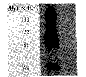
图1 抗人肺癌mAbN-35与肺癌相关蛋白N35的关系
Fig1 Relationship between anti-N35 mAb and associated protein N35 by Western blot
The bands with Mr of 150×103 and 50×103 were separately the light
, http://www.100md.com
chain and heavy chain of mouse IgG.The band with Mr of
75×103-130×103 was the associated protein N35.
2.2 相关蛋白N35在不同组织及细胞中的分布 免疫印迹法测定的结果见图2。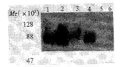
图2 相关蛋白N35在不同组织及细胞中的分布
Fig2 The distribution of associated protein N35 in various
tis-sues and cells by Western blot
, 百拇医药
The lanes of 1,2,3,4,5 and 6 were separately:HeLa,GLC-82,HepG-2,PMC,normal human heart and lung tissues.The associated protein N35 detected
by anti-N35 mAb distributed in GLC-82(Mr≈75×103-130×103),HeLa(Mr≈75×103-130×103),HepG-2(Mr≈90×103-110×103)and
PMC(Mr≈80×103-95×103),but no expression in the protein
ingredient of fresh normal human heart and lung tissues.
, http://www.100md.com
图3 相关蛋白N35在亚细胞结构中的表达
Fig3 The subcellular localization of the associated protein N35
by subcellular fractionation and Westen blot
The lanes of 1,2,3,4 and 5 were separately:cell residue,supernatant from last centrifugation,nuclei,mitochondria and membrane of the cell line GLC-82
harvested from the differential centrifugation.Western blot analysis of
, 百拇医药
subcellular structure[volume(100μg/well)] or protein amount〔(100μg/well)〕
showed the associated protein N35 distributed significantly in the nuclei,distribution in the mitochondrail was a fewer,distribution in menbrane was very few.
2.3 相关蛋白N35在亚细胞结构中的表达 经差速离心法,获得细胞碎片、细胞核成分、线粒体成分和细胞膜成分。在平均蛋白量100μg/孔或平均体积量100μL/孔的条件下,免疫印迹结果显示,相关蛋白N35主要分布于核成分中,线粒体次之,膜成分中含量最少。末次差速离心后的上清中,已无相关蛋白N35可测出,表明经3次差速离心后,细胞裂解液中的有关成分 已完全收集。
, http://www.100md.com
2.4 相关蛋白N35在有丝分裂细胞中的定位 以抗N35mAb对同一视野用不同光源获得的荧光染色摄影图像中,可见有丝分裂细胞中呈蓝染的DNA(赤道板)结构(图4A)。DNA结构的两侧精确对应位置的中心粒处呈现清晰的免疫荧光染色 ,并对称分布于赤道板的两侧(图4B)。
图4 相关蛋白N35在有丝分裂细胞中的定位
Fig4 The localization of the associated protein N35 in
the mi-totic cells by immunoflourescence staining(×400)
The associated protein N35 was located on the centriole of the chromosomeat
, 百拇医药
theS-G2phase.Fig4AandFig4Bwereexposedfromthesamefieldexactlywit hdifferentfluorescencesource.
4A:TheDNA(equatorialplate)wasdyedblue;
4B:TheclearimmunoflourescencestainingoftheassociatedproteinN35presen
tedinpositionofthecentriolesanddistributedsymmetricallyonthebothside softheequatorialplate.
2.5 相关蛋白N35的酶解 GLC-82细胞裂解物用N-glycanase消化(37℃1h)后,再以抗N35mAb进行免疫印迹(图5)。第1泳道可见消化后的相关蛋白N35分解成Mr≈65×103,80×103及 86×1033个不同的小片段;第2泳道可见未经消化的相关蛋白N35(Mr≈75×103~130×103)。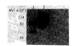
, http://www.100md.com
图5 肺癌相关蛋白N35的酶解
Fig5 The enzyme digestion of the associated protein N35
After lysate of GLC-82 cells wasdigested with N-glycanase,the associated
protein N35 was detected by Western blot with anti-N35 mAb.
Lane1:Digested N35 proteinwas cloven into 3 fragments with Mr of
65×103,84×103 and 86×103;Lane2:Undigested associated N35
, 百拇医药
protein with Mr of 75×103-130×103.
3 讨论
在肿瘤免疫学的研究中,抗肿瘤mAb正在发挥着日益重要的作用〔1-3〕。从临床诊治的意义出发,3种重要的生物大分子靶(即酶、受体和核酸),一直受到很大的重视。近年来由于蛋白质化学技术的飞速发展,一些重要的酶和受体已分离纯化,有的已获得结晶体结构。随之而来的以蛋白质及其编码基因为靶分子的研究,也取得了令人瞩目的结果〔12〕。其中与恶性肿瘤细胞无限增殖行为相关的蛋白分子尤其重要〔13〕。利用抗肿瘤mAb研究和确定相关抗原分子在肿瘤细胞中的分布及有关特性,对于了解这些分子在恶性肿瘤细胞生物学行为中的作用具有十分重要的意义,并在近年来取得长足进展〔2〕。我们利用本所先后建立的GLC-82细胞系和抗相关蛋白分子F19,SIA2和N35的mAb〔3,4〕,研究了抗人肺癌mAb所识别的抗原分子的有关特性,对于了解这些分子在恶性肿瘤细胞生物学行为中的作用具有十分重要的意义〔13〕。我们以抗N-35mAb作为免疫探针,采用免疫沉淀和免疫印迹技术,证实相关蛋白N35的Mr为75×103~130×103,并以不同Mr的形式分布于GLC-82,HeLa,HepG-2和PMC的蛋白组分中;而在正常人心及肺组织蛋白组分中未见表达。真核细胞的糖蛋白分子通常以膜蛋白和跨膜蛋白的形式存在,并多具有受体或通道的功能〔14〕。我们用N-聚糖酶切割后,相关蛋白N35的Mr由86×103~130×103分解成Mr为65×103,84×103及75×1033个不同的小片段。并证实相关蛋白N35是糖蛋白,主要分布在肿瘤细胞的胞核成分,其在亚细胞结构的分布密度依次为胞核→线粒体→胞膜,不支持其以膜蛋白或跨膜蛋白的形式存在。在进行有丝分裂的GLC-82细胞中,该蛋白分子定位于细胞有丝分裂的中、后期的中心粒上。这不仅进一步提示相关蛋白N35主要是一种核蛋白,而且其极可能与肿瘤细胞的增殖密切相关〔6,13〕,并可能具有调节肿瘤细胞生长的作用,值得进一步研究和开发。
, http://www.100md.com
基金项目:国家自然科学基金资助,No.39960078; 云南省九五重点学科肿瘤学经费资助
作者简介:王秦秦,男,50岁,副主任医师昆明市西昌路153号,Tel.(0871)5324888-2777 Email:zys@public.km.yn.cn2000byJCellMolImmunolPress www.immunology.net/jcmi;Email.immuedit@fmmu.edu.cn
参考文献:
〔1〕 Cao Y, Christian S, Suresh MR, et al. Development of a bispecific monoclonal antibody as a universal immunoprobe for detecting biotinylated macromolecules〔J〕 . J Immunol Meth, 1998;220(1- 2):85- 91.
, 百拇医药
〔2〕 Subong EN, Shue MJ, Epstein JI, et al. Monoclonal antibody to prostate cancer nuclear matrix protein (PRO:4- 216) recognizes nucleophosmin/B23〔J〕 . Prostate, 1999;39(4):298- 304.
〔3〕 Kato S, Ishihara K, Shinozawa T, et al. Monoclonal antibody to human midkine reveasl increased midkine expression in human brain tumors〔J〕 . J Neuropathol Exp Neurol,1999;58(5):430- 441.
〔4〕 梁明达 ,胡美英,贾伟 ,等 .云南个旧肺腺癌细胞系GLC82的建立及特性分析〔J〕. 中华肿瘤杂志, 1985; 7( 2) : 81- 83.
, 百拇医药
〔5〕王秦秦,郝萍 ,孟桦 ,等 . 抗人肺癌单克隆抗体的研制及免疫组化分析43例 〔J〕 . 中华肿瘤杂志 , 1992; 14( 6) : 473.
〔6〕 Yeo JP, Alderuccio F, Toh BH, et al. A new chromosomal protein essential for mitotic spindle assembly〔 J〕.Nature,1994;386:488- 491.
〔7〕 Bobinnec Y, Khodjakov A, Mir LM, et al. Centriole disassembly in vivo and its effect on centrosome structure and function in vertebrate cells〔 J〕 . J Cell Biol, 1998; 143(14):1575- 1589.
, 百拇医药
〔8〕 Domloge- Hultsch N, Utecht L, James W, et al. Autoantibodies from patients with localized and generalized bullous pemphigoid immunoprecipitate the same 230- kd keratinocyte antigen〔 J〕 . Arch Dermatol, 1990; 126(10):1337- 1340.
〔9〕 Weber MM, Spottl G, Gossl C, et al. Characterization of human insulin- like growth factor binding proteins by two- dimensional polyacrylamide gel electrophoresis and Western blot analysis〔 J〕 . J Clin Endocrinol Metab,1999; 84(5):1679- 1684.
, http://www.100md.com
〔10〕 Tomas JE, Smith M, Rubinfeld B, et al. Subcellular localization and analysis of apparent 180 kDa and 220kDa proteins of the breast cacer susceptibility gene, BRCA1〔 J〕 . J Biol Chem, 1996;271(45):28630- 28635.
〔11〕 Ghosh S, Paweletz N, Schroeter D, et al. Effects of okadaic acid on mitotic HeLa cells〔 J〕 . J Cell Sci,.1992;103(Pt 1):117- 124.
〔12〕 Yonezawa N, Fukui N, Kudo K, et al. Localization of neutral N- linked carbohydrate chains in pig zona pellucida glycoprotein〔 J〕 . J Immunol, 1999; 49(5):515- 522.
, http://www.100md.com
〔13〕杨铭 . 以 DNA为靶的小分子药物研究中的分子识别 〔J〕. 北京医科大学学报,1998;
30(2):96- 98.
〔14〕 Levine DS, Sanchez CA, Rabinovitch PS, et al. Formation of the tetraploid intermediate is associated with the development of cells with more than four centrioles in the elastase- simian virus 40 tumor antigen transgenic mouse
model of pancreatic cancer〔 J〕 . Proc Natl Acad Sci USA, 1991; 88(15):6427- 6431.
〔15〕 陈诗书,汤雪明,王一飞,等编著 . 医学细胞与分子生物学〔M〕 .上海: 上海医科大学出版社 , 1996: 316.
收稿日期:1999-07-15
修回日期:1999-10-10, 百拇医药