阻塞性睡眠呼吸暂停综合征患者上气道大小的磁共振研究
作者:高雪梅 曾祥龙 傅民魁 黄席珍 陈雷
单位:高雪梅 曾祥龙 傅民魁 北京医科大学口腔医学院,北京 100081黄席珍 北京协和医院;陈雷 人民医院CT磁共振中心
关键词:睡眠呼吸暂停综合征;病理学;咽;解剖学和组织学;咽;放射性核素成像;核磁共振
北京医科大学学报990519 摘 要 目的:以软组织显影好的磁共振技术(magnetic resonance imaging,MRI)探讨阻塞性睡眠呼吸暂停综合征(obstructive sleep apnea syndrome, OSAS)患者上气道与无鼾正常人的差异。方法:23名OSAS患者均经整夜多导睡眠监测确诊。每2或1名OSAS患者选取1名年龄、性别配比的无鼾对照者。对23名OSAS患者与12名配对对照的上气道进行MRI扫描及测量,比较鼻咽、腭咽、舌咽和喉咽的大小。结果:OSAS患者的上气道各段,在矢向径、横向径、矢向/横向径比、截面积和体积几乎各项指标上均普遍小于无鼾对照,其鼻咽尤其值得重视。结论:OSAS存在形态学病因机制。MRI在OSAS研究中是有重要价值的诊断工具,但不能替代整夜睡眠监测。
, http://www.100md.com
中国图书资料分类法分类号 R766.5
The magnetic resonance imaging of the upper airway
in obstructive sleep apnea syndrome
GAO Xue-Mei#, ZENG Xiang-Long, FU Min-Kui, HUANG Xi-Zhen, CHEN Lei
(#Department of Orthodontics, School of Stamatology, Beijing Medical University , Beijing 100081 China)
MeSH Sleep apnea syndromes/pathol Pharynx/anat Pharynx/radionaclide Nuclear magnetic resonance
, http://www.100md.com
ABSTRACT Objective: We used magnetic resonance imaging(MRI) which could provide super imaging of soft tissue to investigate the difference between the obstructive sleep apnea syndrome(OSAS) and the non-snorers. Methods: 23 OSAS patients were diagnosed by whole night polysomnography. Every one or two patients had one age-sex-matched control subject with no snoring at night. Both 23 OSAS patients and 12 controls took MRI scans of upper airway. Compared those images within nasopharynx, palatopharynx, glossopharynx and hypopharynx. Results: OSAS patients showed smaller airway than controls in almost every aspect, such as sagittal size, horizontal size, the ratio of sagittal size to horizontal size, cross-sectional area and volume of the upper airway. The difference in the nasopharynx was an interesting discovery. Conclusion: A structurally abnormal airway might serve as an anatomic substrate for the development of sleep apnea. MRI was a valuable diagnostic tool in the research of OSAS, though it could not be replaced of whole night polysomnography.
, http://www.100md.com
(J Beijing Med Univ,1999,31:450-453)
阻塞性睡眠呼吸暂停综合征(obstructive sleep apnea syndrome,OSAS)为夜间反复发生的上气道狭窄或阻塞,近年来日益受到研究者重视。不规则的上气道,只有通过三维手段才能正确认识其形态。自1983年Suratt[1]起,一些学者以CT(computeri-
zed tomography)为手段进行了OSAS形态学研究,迅速丰富了人们对OSAS的认识,但存在不足:(1)均是CT研究,对软组织显影较差;(2)样本量较小,测量指标也较少;(3)对照组在性别、年龄等因素上配对不严格。国内有刘月华[2]等做OSAS的X线和CT影像学分析。作为软组织显影好的核磁共振影像技术(magnetic resonance imaging, MRI)研究,甚少用于OSAS上气道一般特征的研究。
, 百拇医药
1 对象与方法
1.1 对象
研究组23名OSAS患者均为1996~1997年的我科门诊患者。其中男性20人,女性3人。所有病人经协和医院夜间多导睡眠图确诊为符合标准的OSAS[3]。其呼吸暂停低通气指数(apnea/hypopnea index, AHI)为(45±24)次.h-1, 从5.82次.h-1至94.60次.h-1。
配对对照组来自40岁以上无OSAS症状及其相关疾病的北京医科大学人民医院磁共振中心的门诊患者,每位对照与1~2位相近年龄性别的OSAS相对应。共选取12人,其中男10人,女2人。无睡眠呼吸障碍判定标准: (1)自述或被家人证实无持续/响亮鼾声;(2)无夜间憋气或被发现夜间呼吸暂停现象;(3)无晨起头痛、恶心等症状及日间严重困倦;(4)无阻塞性肺疾病及上气道肿物或压迫气道的疾病,鼻腔通畅;(5)无影响呼吸中枢的疾病,如脑干肿瘤或梗塞等;(6)无甲状腺功能低下、垂体瘤等疾病。此组均未做夜间多导睡眠监测。
, http://www.100md.com
以上两组人员在性别构成、年龄、身高、体重上差异均无显著性,体重指数BMI差异有显著性(表1)。
表1 OSAS研究组与配对对照组的一般情况的差异比较( ±s)
±s)
Table 1 Clinical characteristics of OSAS patients and those of the controls( ±s) Group
Matched controls
OSAS
n(male/female)
, http://www.100md.com
10/2
20/3
Age(years)
49.8±10.1
50.3±10.9
Height(cm)
171.8± 6.8
168.2± 5.6
Weight(kg)
71.3± 7.6
75.7± 8.7
BMI(kg.m-2)
, http://www.100md.com
24.1± 1.9
26.8± 3.0**
AHI(episodes/h-1)
/
45.7±24.0
** P<0.01, compared with the matched controls.1.2 方法
研究组及无睡眠呼吸障碍组每一成员均扫描MRI。投照机器为北京医科大学人民医院磁共振中心的MRI,机型GYREX-2T SGR(以色列产ELSCint Ltd.)。头线圈。SE序列T1加权像,TR/TE=620-680/12.0,层厚5 mm,间隔1.5 mm。取像范围:轴位(包含自鼻咽顶端至第三颈椎水平),矢状位(下颌骨内缘范围)。距阵256×256,激励次数2次。
, http://www.100md.com
研究组及无睡眠呼吸障碍组成员均以仰卧位扫描,眼耳平面垂直于地,正中合,平静呼吸,扫描中嘱勿吞咽及讲话,并保持清醒。扫描数据存入可擦写光盘(3M公司,1.2G)。
在Elscint工作站上,输入扫描数据,固定窗位C1=-732,固定窗宽W1=665。利用计算机固化程序描选所要测定的面积,读取上气道轴位截面面积、舌体及软腭矢状截面面积,测取3遍,最后计算取其平均值;同样利用工作站固化软件测量咽腔直径,矢状径为轴截面上通过头颅矢状轴的线段,横径为与矢状径成90度角的线段。
1.3 MRI测量项目及统计学处理
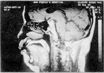
在正中矢状面的计划线定位图上判定咽腔各层次(图1)。
The nasopharynx was defined as the region between the roof of the airway and the hard palate. The palatopharynx lies between the hard palate and the tip of the soft palate. The glossopharynx was from the tip of the soft palate to the top of the epiglottis. The hypopharynx is between the top of the epiglottis and the base of epiglottis. All 19 images of upper airway could be divided into these four major levels.
, 百拇医药
图1 MRI鼻咽、腭咽、舌咽、喉咽的划分
Figure 1 The division of upper airway: nasopharynx,palatopharynx, glossopharynx and hypopharynx
记录和计算咽腔各段内最小截面积、最大截面积、平均截面积;平均矢状直径、平均横向直径;矢向径与横向径的比例;按层厚5 mm、间隔1.5 mm累积计算咽腔各段的体积。在计算机上利用SPSS(Window版)统计软件包进行各计量资料t检验。
2 结果
研究组(OSAS患者)与配对对照组上气道直径的比较见表2。
表2 OSAS与配对对照上气道MRI直径比较( ±s)
±s)
, http://www.100md.com
Table 2 The upper airway size (mm) of OSAS compared with the matched controls( ±s)
±s)
d/mm
Sagittal size
Horizontal size
Matched controls
OSAS
Matched controls
OSAS
Nasopharynx
, 百拇医药
17.8±2.4
14.3±2.8△△△
24.8±2.3
22.6±3.1△
Palatopharynx
8.2±2.9
7.1±2.6
15.0±5.1
13.0±3.9
Glossopharynx
11.9±1.7
, 百拇医药
9.3±3.2△△
24.6±8.7
17.7±5.8△△
Hypopharynx
14.8±4.7
11.4±2.8△
28.3±7.2
26.2±7.5
△P<0.05, △△P<0.01, △△△P<0.001, compared with matched controls. 研究组(OSAS病人)与配对对照组上气道矢向/横向径比的比较见表3。
, http://www.100md.com
表3 OSAS与配对对照上气道矢状/横向径比( ±s)
±s)
Table 3 The ratio of sagittal size to horizontal size of upper airway in OSAS compared with the matched controls( ±s) Group
Max
Min
Mean
Control
OSAS
, 百拇医药
Control
OSAS
Control
OSAS
Nasopharynx
0.84±0.15
0.73±0.15△
0.61±0.10
0.55±0.18
0.72±0.10
0.64±0.13
Palatopharynx
, 百拇医药
0.80±0.27
0.87±0.34
0.35±0.20
0.36±0.26
0.57±0.21
0.59±0.27
Glossopharynx
0.64±0.22
0.89±0.91
0.43±0.18
0.46±0.32
0.53±0.18
, 百拇医药
0.63±0.52
Hypopharynx
0.68±0.19
0.64±0.26
0.43±0.11
0.33±0.19
0.53±0.13
0.47±0.19
△P<0.05, compared with the matched controls. 研究组(OSAS病人)与配对对照组上气道截面积的比较见表4、图2。
表4 OSAS与配对对照上气道MRI截面积比较( ±s)
±s)
, http://www.100md.com
Table 4 The section area(mm2) of upper airway in OSAS compared with the matched controls( ±s)
±s)
A/mm2 Group
Mean
Min
Control
OSAS
Control
OSAS
Nasopharynx
, 百拇医药
363±52
269±75△△△
315±63
232±84△△
Palatopharynx
121±72
85±38
56±24
29±14△△
Glossopharynx
194±71
, 百拇医药
115±46△△△
152±79
75±38△△
Hypopharynx
244±107
176±67△
191±119
114±72
△P<0.05, △△P<0.01, △△△P<0.001, compared with matched controls.

, 百拇医药
Upper, a male OSAS patients; bottom, a matched control1
图2 OSAS患者与配对对照的上气道比较
Figure 2 The narrow upper airway of OSAS compared with that of the matched control
研究组(OSAS病人)与配对对照组上气道体积的比较见表5。
表5 OSAS与配对对照上气道及周围结构体积比较( ±s)
±s)
Table 5 The volume of upper airway and surrounding tissues in OSAS compared with the matched controls( ±s)
±s)
, 百拇医药
V/mm3 Group
Control
OSAS
nasopharynx
5 321±1 525
3 589±1 336△△
Palatopharynx
4 018±3 526
2 560±1 281
Glossopharynx
4 982±2 917
, 百拇医药
3 167±1 525△
hypopharynx
4 868±1 499
3 706±1 793
Oropharynx
9 000±6 241
5 727±2 160
Whole pharynx
19 189±6 241
13 022±4 277△△
Soft palate
, http://www.100md.com
6 794±2 208
7 556±2 079
Tongue
87 455±11 891
99 390±16 053△
△P<0.05, △△P<0.01, compared with matched controls. 3 讨论
3.1 MRI是上气道形态研究极有价值的手段
MRI具有软组织显影好,无放射侵害,任意截面等优点,可更好地观察上气道的形态[4,9]。但是由于MRI造价较高,既往研究中甚少见到用于OSAS形态的报告。本研究使用MRI先进影像手段,观察样本量于同类研究中较大,对照选择适当。
, 百拇医药
3.2 OSAS的形态特点
总体上OSAS在上气道各个区段均表现出狭窄,40%的指标与配对对照有显著差异,如果增加样板量,可能有更多有统计学意义的显著性表现。这与其他学者通过声音反射技术、CT以及X线研究的相关研究结论一致[1,5-7]。这说明上气道形态在OSAS的发病中占有一席之地。 本研究涉及上气道的阻塞部位。OSAS上气道存在一个或几个阻塞部位,通常发生在最小截面积处,这一点已通过夜间X线透视得到证实[1]。所以长期以来最小截面积一直被许多学者作为研究阻塞的指标[1,5]。在本研究中,腭咽最小截面积于OSAS组和对照组中差异没有统计学意义,提示腭咽最狭窄点的大小不是OSAS的特征指标;而舌咽的最小截面积在两组间有显著性差异,则提示孰能扩大舌咽,孰能获得治疗OSAS较高的成功率。腭咽成形术的有效率大约只有50%,可能与该手术只针对腭咽有关。口腔矫正器能够作用到腭咽和舌咽,所以有效率可以达到80%左右。
, 百拇医药
本研究看到OSAS患者鼻咽与对照组比,差异的广泛性和显著性均大,值得重视。因为阻塞较少观察到发生在鼻咽,普遍对OSAS的鼻咽没引起足够的重视。而本研究则感到,有必要强调OSAS的鼻咽,鼻咽更少软组织代偿的干扰,且可能揭示特殊的OSAS上气道动力学病因[8]。
3.3 OSAS患者上气道形态研究的意义
众多学者致力于快捷高效能的上气道影像技术的研究,希望以此代替冗长繁琐的整夜睡眠监测。有学者提出一些指标,如软腭形态[6]、咽后壁厚度[7]、上气道最小截面积[5]等,但作为诊断,其特异性和敏感性多不足以达到指标要求。
本研究从鼻咽至喉咽,尽可能涵盖了可变(无软骨包绕)上气道的全程;每一层面包括线距、线距比、面积和体积,试图筛选出特异性诊断指标。但是各项上气道测量值标准差都较大,研究组与对照组之间的交叉重叠较多,说明上气道在各个体之间差异较大,单独评判某个患者的上气道是大是小没有意义,影像仍然尚不足以作为独立的诊断手段。
, 百拇医药
OSAS毕竟是一个与形态紧密相关的疾病,有待我们更新技术手段,寻找更敏感指标,进一步增强影像在诊断上的作用。
(本研究得到我校人民医院杜湘柯、高健、张桂青、齐净及第一医院高峰同志的大力帮助,特此致谢!)
注:国家自然科学基金(39670791)基金项目。
参考文献
1 Suratt PM, Dee P, Atkinson RL, et al. Fluoroscopic and computed tomographic features of the pharyngeal airway in obstructive sleep apnea. Am Rev Respir Dis, 1983, 127:487-492
2 刘月华,曾祥龙,傅民魁,等.阻塞性睡眠呼吸暂停综合征与上气道及颜面结构的相关研究.中华医学杂志, 1998,78:849
, 百拇医药
3 黄席珍,吴全有,李龙云,等.多导睡眠图的临床应用.中华内科杂志, 1991,30:258-261
4 Ryan CF, Lowe AA, Li D, et al. Magnetic resonance imaging of the upper airway in obstructive sleep apnea before and after chronic nasal continuous positive airway pressure therapy. Am Rev Respir Dis, 1991;144:939-944
5 Avrahami E, Englender M. Relation between axial cross-sectional area of the oropharynx and obstructive sleep apnea syndrome in adult. AJNR Am J Neuroradiol, 1995, 16: 135-140
, 百拇医药
6 Pepin JL, Veale D, Ferretti GR, et al. Obstructive sleep apnea syndrome:hoooked appearance of the soft palate in awake patients-cephalometric and CT findings. Radiology, 1999;210:163-170
7 Caballero P, Alvarez SR, Garcia GF, et al. CT in the evaluation of the upper airway in healthy subjects and in patients with obstructive sleep apnea syndrome. Chest, 1998,113:111-116
8 高雪梅,曾祥龙,傅民魁,等.鼻咽大小对OSAS病人的影响.中华耳鼻咽喉科杂志, 1999,34:166-169
9 Hsegawa K, Nishimura T, Yagisawa M, et al. Diagnosis by dynamic MRI in sleep disordered breathing. Acta Otolaryngol Suppl Stockh, 1996,523:245-247
(1998-11-04收稿), http://www.100md.com
单位:高雪梅 曾祥龙 傅民魁 北京医科大学口腔医学院,北京 100081黄席珍 北京协和医院;陈雷 人民医院CT磁共振中心
关键词:睡眠呼吸暂停综合征;病理学;咽;解剖学和组织学;咽;放射性核素成像;核磁共振
北京医科大学学报990519 摘 要 目的:以软组织显影好的磁共振技术(magnetic resonance imaging,MRI)探讨阻塞性睡眠呼吸暂停综合征(obstructive sleep apnea syndrome, OSAS)患者上气道与无鼾正常人的差异。方法:23名OSAS患者均经整夜多导睡眠监测确诊。每2或1名OSAS患者选取1名年龄、性别配比的无鼾对照者。对23名OSAS患者与12名配对对照的上气道进行MRI扫描及测量,比较鼻咽、腭咽、舌咽和喉咽的大小。结果:OSAS患者的上气道各段,在矢向径、横向径、矢向/横向径比、截面积和体积几乎各项指标上均普遍小于无鼾对照,其鼻咽尤其值得重视。结论:OSAS存在形态学病因机制。MRI在OSAS研究中是有重要价值的诊断工具,但不能替代整夜睡眠监测。
, http://www.100md.com
中国图书资料分类法分类号 R766.5
The magnetic resonance imaging of the upper airway
in obstructive sleep apnea syndrome
GAO Xue-Mei#, ZENG Xiang-Long, FU Min-Kui, HUANG Xi-Zhen, CHEN Lei
(#Department of Orthodontics, School of Stamatology, Beijing Medical University , Beijing 100081 China)
MeSH Sleep apnea syndromes/pathol Pharynx/anat Pharynx/radionaclide Nuclear magnetic resonance
, http://www.100md.com
ABSTRACT Objective: We used magnetic resonance imaging(MRI) which could provide super imaging of soft tissue to investigate the difference between the obstructive sleep apnea syndrome(OSAS) and the non-snorers. Methods: 23 OSAS patients were diagnosed by whole night polysomnography. Every one or two patients had one age-sex-matched control subject with no snoring at night. Both 23 OSAS patients and 12 controls took MRI scans of upper airway. Compared those images within nasopharynx, palatopharynx, glossopharynx and hypopharynx. Results: OSAS patients showed smaller airway than controls in almost every aspect, such as sagittal size, horizontal size, the ratio of sagittal size to horizontal size, cross-sectional area and volume of the upper airway. The difference in the nasopharynx was an interesting discovery. Conclusion: A structurally abnormal airway might serve as an anatomic substrate for the development of sleep apnea. MRI was a valuable diagnostic tool in the research of OSAS, though it could not be replaced of whole night polysomnography.
, http://www.100md.com
(J Beijing Med Univ,1999,31:450-453)
阻塞性睡眠呼吸暂停综合征(obstructive sleep apnea syndrome,OSAS)为夜间反复发生的上气道狭窄或阻塞,近年来日益受到研究者重视。不规则的上气道,只有通过三维手段才能正确认识其形态。自1983年Suratt[1]起,一些学者以CT(computeri-
zed tomography)为手段进行了OSAS形态学研究,迅速丰富了人们对OSAS的认识,但存在不足:(1)均是CT研究,对软组织显影较差;(2)样本量较小,测量指标也较少;(3)对照组在性别、年龄等因素上配对不严格。国内有刘月华[2]等做OSAS的X线和CT影像学分析。作为软组织显影好的核磁共振影像技术(magnetic resonance imaging, MRI)研究,甚少用于OSAS上气道一般特征的研究。
, 百拇医药
1 对象与方法
1.1 对象
研究组23名OSAS患者均为1996~1997年的我科门诊患者。其中男性20人,女性3人。所有病人经协和医院夜间多导睡眠图确诊为符合标准的OSAS[3]。其呼吸暂停低通气指数(apnea/hypopnea index, AHI)为(45±24)次.h-1, 从5.82次.h-1至94.60次.h-1。
配对对照组来自40岁以上无OSAS症状及其相关疾病的北京医科大学人民医院磁共振中心的门诊患者,每位对照与1~2位相近年龄性别的OSAS相对应。共选取12人,其中男10人,女2人。无睡眠呼吸障碍判定标准: (1)自述或被家人证实无持续/响亮鼾声;(2)无夜间憋气或被发现夜间呼吸暂停现象;(3)无晨起头痛、恶心等症状及日间严重困倦;(4)无阻塞性肺疾病及上气道肿物或压迫气道的疾病,鼻腔通畅;(5)无影响呼吸中枢的疾病,如脑干肿瘤或梗塞等;(6)无甲状腺功能低下、垂体瘤等疾病。此组均未做夜间多导睡眠监测。
, http://www.100md.com
以上两组人员在性别构成、年龄、身高、体重上差异均无显著性,体重指数BMI差异有显著性(表1)。
表1 OSAS研究组与配对对照组的一般情况的差异比较(
 ±s)
±s)Table 1 Clinical characteristics of OSAS patients and those of the controls(
 ±s) Group
Matched controls
OSAS
n(male/female)
, http://www.100md.com
10/2
20/3
Age(years)
49.8±10.1
50.3±10.9
Height(cm)
171.8± 6.8
168.2± 5.6
Weight(kg)
71.3± 7.6
75.7± 8.7
BMI(kg.m-2)
, http://www.100md.com
24.1± 1.9
26.8± 3.0**
AHI(episodes/h-1)
/
45.7±24.0
** P<0.01, compared with the matched controls.1.2 方法
研究组及无睡眠呼吸障碍组每一成员均扫描MRI。投照机器为北京医科大学人民医院磁共振中心的MRI,机型GYREX-2T SGR(以色列产ELSCint Ltd.)。头线圈。SE序列T1加权像,TR/TE=620-680/12.0,层厚5 mm,间隔1.5 mm。取像范围:轴位(包含自鼻咽顶端至第三颈椎水平),矢状位(下颌骨内缘范围)。距阵256×256,激励次数2次。
, http://www.100md.com
研究组及无睡眠呼吸障碍组成员均以仰卧位扫描,眼耳平面垂直于地,正中合,平静呼吸,扫描中嘱勿吞咽及讲话,并保持清醒。扫描数据存入可擦写光盘(3M公司,1.2G)。
在Elscint工作站上,输入扫描数据,固定窗位C1=-732,固定窗宽W1=665。利用计算机固化程序描选所要测定的面积,读取上气道轴位截面面积、舌体及软腭矢状截面面积,测取3遍,最后计算取其平均值;同样利用工作站固化软件测量咽腔直径,矢状径为轴截面上通过头颅矢状轴的线段,横径为与矢状径成90度角的线段。
1.3 MRI测量项目及统计学处理
在正中矢状面的计划线定位图上判定咽腔各层次(图1)。

The nasopharynx was defined as the region between the roof of the airway and the hard palate. The palatopharynx lies between the hard palate and the tip of the soft palate. The glossopharynx was from the tip of the soft palate to the top of the epiglottis. The hypopharynx is between the top of the epiglottis and the base of epiglottis. All 19 images of upper airway could be divided into these four major levels.
, 百拇医药
图1 MRI鼻咽、腭咽、舌咽、喉咽的划分
Figure 1 The division of upper airway: nasopharynx,palatopharynx, glossopharynx and hypopharynx
记录和计算咽腔各段内最小截面积、最大截面积、平均截面积;平均矢状直径、平均横向直径;矢向径与横向径的比例;按层厚5 mm、间隔1.5 mm累积计算咽腔各段的体积。在计算机上利用SPSS(Window版)统计软件包进行各计量资料t检验。
2 结果
研究组(OSAS患者)与配对对照组上气道直径的比较见表2。
表2 OSAS与配对对照上气道MRI直径比较(
 ±s)
±s), http://www.100md.com
Table 2 The upper airway size (mm) of OSAS compared with the matched controls(
 ±s)
±s)d/mm
Sagittal size
Horizontal size
Matched controls
OSAS
Matched controls
OSAS
Nasopharynx
, 百拇医药
17.8±2.4
14.3±2.8△△△
24.8±2.3
22.6±3.1△
Palatopharynx
8.2±2.9
7.1±2.6
15.0±5.1
13.0±3.9
Glossopharynx
11.9±1.7
, 百拇医药
9.3±3.2△△
24.6±8.7
17.7±5.8△△
Hypopharynx
14.8±4.7
11.4±2.8△
28.3±7.2
26.2±7.5
△P<0.05, △△P<0.01, △△△P<0.001, compared with matched controls. 研究组(OSAS病人)与配对对照组上气道矢向/横向径比的比较见表3。
, http://www.100md.com
表3 OSAS与配对对照上气道矢状/横向径比(
 ±s)
±s)Table 3 The ratio of sagittal size to horizontal size of upper airway in OSAS compared with the matched controls(
 ±s) Group
Max
Min
Mean
Control
OSAS
, 百拇医药
Control
OSAS
Control
OSAS
Nasopharynx
0.84±0.15
0.73±0.15△
0.61±0.10
0.55±0.18
0.72±0.10
0.64±0.13
Palatopharynx
, 百拇医药
0.80±0.27
0.87±0.34
0.35±0.20
0.36±0.26
0.57±0.21
0.59±0.27
Glossopharynx
0.64±0.22
0.89±0.91
0.43±0.18
0.46±0.32
0.53±0.18
, 百拇医药
0.63±0.52
Hypopharynx
0.68±0.19
0.64±0.26
0.43±0.11
0.33±0.19
0.53±0.13
0.47±0.19
△P<0.05, compared with the matched controls. 研究组(OSAS病人)与配对对照组上气道截面积的比较见表4、图2。
表4 OSAS与配对对照上气道MRI截面积比较(
 ±s)
±s), http://www.100md.com
Table 4 The section area(mm2) of upper airway in OSAS compared with the matched controls(
 ±s)
±s)A/mm2 Group
Mean
Min
Control
OSAS
Control
OSAS
Nasopharynx
, 百拇医药
363±52
269±75△△△
315±63
232±84△△
Palatopharynx
121±72
85±38
56±24
29±14△△
Glossopharynx
194±71
, 百拇医药
115±46△△△
152±79
75±38△△
Hypopharynx
244±107
176±67△
191±119
114±72
△P<0.05, △△P<0.01, △△△P<0.001, compared with matched controls.


, 百拇医药
Upper, a male OSAS patients; bottom, a matched control1
图2 OSAS患者与配对对照的上气道比较
Figure 2 The narrow upper airway of OSAS compared with that of the matched control
研究组(OSAS病人)与配对对照组上气道体积的比较见表5。
表5 OSAS与配对对照上气道及周围结构体积比较(
 ±s)
±s)Table 5 The volume of upper airway and surrounding tissues in OSAS compared with the matched controls(
 ±s)
±s), 百拇医药
V/mm3 Group
Control
OSAS
nasopharynx
5 321±1 525
3 589±1 336△△
Palatopharynx
4 018±3 526
2 560±1 281
Glossopharynx
4 982±2 917
, 百拇医药
3 167±1 525△
hypopharynx
4 868±1 499
3 706±1 793
Oropharynx
9 000±6 241
5 727±2 160
Whole pharynx
19 189±6 241
13 022±4 277△△
Soft palate
, http://www.100md.com
6 794±2 208
7 556±2 079
Tongue
87 455±11 891
99 390±16 053△
△P<0.05, △△P<0.01, compared with matched controls. 3 讨论
3.1 MRI是上气道形态研究极有价值的手段
MRI具有软组织显影好,无放射侵害,任意截面等优点,可更好地观察上气道的形态[4,9]。但是由于MRI造价较高,既往研究中甚少见到用于OSAS形态的报告。本研究使用MRI先进影像手段,观察样本量于同类研究中较大,对照选择适当。
, 百拇医药
3.2 OSAS的形态特点
总体上OSAS在上气道各个区段均表现出狭窄,40%的指标与配对对照有显著差异,如果增加样板量,可能有更多有统计学意义的显著性表现。这与其他学者通过声音反射技术、CT以及X线研究的相关研究结论一致[1,5-7]。这说明上气道形态在OSAS的发病中占有一席之地。 本研究涉及上气道的阻塞部位。OSAS上气道存在一个或几个阻塞部位,通常发生在最小截面积处,这一点已通过夜间X线透视得到证实[1]。所以长期以来最小截面积一直被许多学者作为研究阻塞的指标[1,5]。在本研究中,腭咽最小截面积于OSAS组和对照组中差异没有统计学意义,提示腭咽最狭窄点的大小不是OSAS的特征指标;而舌咽的最小截面积在两组间有显著性差异,则提示孰能扩大舌咽,孰能获得治疗OSAS较高的成功率。腭咽成形术的有效率大约只有50%,可能与该手术只针对腭咽有关。口腔矫正器能够作用到腭咽和舌咽,所以有效率可以达到80%左右。
, 百拇医药
本研究看到OSAS患者鼻咽与对照组比,差异的广泛性和显著性均大,值得重视。因为阻塞较少观察到发生在鼻咽,普遍对OSAS的鼻咽没引起足够的重视。而本研究则感到,有必要强调OSAS的鼻咽,鼻咽更少软组织代偿的干扰,且可能揭示特殊的OSAS上气道动力学病因[8]。
3.3 OSAS患者上气道形态研究的意义
众多学者致力于快捷高效能的上气道影像技术的研究,希望以此代替冗长繁琐的整夜睡眠监测。有学者提出一些指标,如软腭形态[6]、咽后壁厚度[7]、上气道最小截面积[5]等,但作为诊断,其特异性和敏感性多不足以达到指标要求。
本研究从鼻咽至喉咽,尽可能涵盖了可变(无软骨包绕)上气道的全程;每一层面包括线距、线距比、面积和体积,试图筛选出特异性诊断指标。但是各项上气道测量值标准差都较大,研究组与对照组之间的交叉重叠较多,说明上气道在各个体之间差异较大,单独评判某个患者的上气道是大是小没有意义,影像仍然尚不足以作为独立的诊断手段。
, 百拇医药
OSAS毕竟是一个与形态紧密相关的疾病,有待我们更新技术手段,寻找更敏感指标,进一步增强影像在诊断上的作用。
(本研究得到我校人民医院杜湘柯、高健、张桂青、齐净及第一医院高峰同志的大力帮助,特此致谢!)
注:国家自然科学基金(39670791)基金项目。
参考文献
1 Suratt PM, Dee P, Atkinson RL, et al. Fluoroscopic and computed tomographic features of the pharyngeal airway in obstructive sleep apnea. Am Rev Respir Dis, 1983, 127:487-492
2 刘月华,曾祥龙,傅民魁,等.阻塞性睡眠呼吸暂停综合征与上气道及颜面结构的相关研究.中华医学杂志, 1998,78:849
, 百拇医药
3 黄席珍,吴全有,李龙云,等.多导睡眠图的临床应用.中华内科杂志, 1991,30:258-261
4 Ryan CF, Lowe AA, Li D, et al. Magnetic resonance imaging of the upper airway in obstructive sleep apnea before and after chronic nasal continuous positive airway pressure therapy. Am Rev Respir Dis, 1991;144:939-944
5 Avrahami E, Englender M. Relation between axial cross-sectional area of the oropharynx and obstructive sleep apnea syndrome in adult. AJNR Am J Neuroradiol, 1995, 16: 135-140
, 百拇医药
6 Pepin JL, Veale D, Ferretti GR, et al. Obstructive sleep apnea syndrome:hoooked appearance of the soft palate in awake patients-cephalometric and CT findings. Radiology, 1999;210:163-170
7 Caballero P, Alvarez SR, Garcia GF, et al. CT in the evaluation of the upper airway in healthy subjects and in patients with obstructive sleep apnea syndrome. Chest, 1998,113:111-116
8 高雪梅,曾祥龙,傅民魁,等.鼻咽大小对OSAS病人的影响.中华耳鼻咽喉科杂志, 1999,34:166-169
9 Hsegawa K, Nishimura T, Yagisawa M, et al. Diagnosis by dynamic MRI in sleep disordered breathing. Acta Otolaryngol Suppl Stockh, 1996,523:245-247
(1998-11-04收稿), http://www.100md.com