水稻条纹病毒病害特异蛋白的提纯及血清学特性
作者:林含新 林奇田 吴祖建 林丽明 林奇英 谢联辉
单位:(福建农业大学植物病毒研究所,福建省植物病毒学重点实验室,福州350002)
关键词:水稻条纹病毒;病害特异蛋白;提纯;血清学特性
中国病毒学/990306 摘 要 应用差别pH值沉淀蛋白质的原理,建立了水稻条纹病毒病特异蛋白(SP)的两种提纯方法。这两种方法都可以从病叶中提纯到大量的SP,其粗提纯量分别为0.8和2.0mg/g病叶。通过SDS-PAGE分离后得到了精提纯的蛋白,其分子量为20.1kDa。将粗提纯和精提纯的SP分别免疫兔子,制备出效价为51200和6400的抗血清。将效价为6400的高度特异性的抗血清用于研究RSV-SP与RSV-CP及同属的水稻草状矮化病毒(RGSV)SP、CP之间的血清学关系,结果表明,RSV-SP的抗血清与RGSV-CP、RSV-CP之间无反应,但可与RGSV-SP微弱反应;而RGSV-SP、CP及RSV-CP的抗血清与RSV-SP之间都无血清学反应。结果证实了RSV和RGSV之间存在着进化上的亲缘关系。
, 百拇医药
Purification and Serological Characteristics of Disease-Specific
Protein of Rice Stripe Virus
Lin Hanxin Lin Qitian Wu Zujian
Lin Liming Lin Qiying Xie Lianhui
(Institute of Plant Virology of Fujian Agricultural University, Key Laboratory
of Plant Virology of Fujian Province, Fuzhou 350002)
, http://www.100md.com
Abstract According to the differential pH precipitation of proteins, two methods were established for purification of disease-specific protein (SP) in rice plants infected by rice stripe virus (RSV), a member of Tenuivirus. The yields obtained by the two methods were 0.8 and 2.0 mg SP per g of infected tissues, respectively. After separation by SDS-PAGE, further purified SP was obtained. The Mr of SP is 20.1 kDa. Antisera against the crude and further purified SP were prepared with the titer of 51 200 and 6 400, respectively. The antiserum against the further purified SP was proved to be highly specific, with which the serological relationships between RSV-SP and RSV-CP, CP and SP of rice grassy stunt virus (RGSV), another member of Tenuivirus were studied. Using PAS-ELISA, it was found that RSV-SP antiserum did not react with RSV-CP and RGSV-CP, but weakly reacted with RGSV-SP. No serological relationship existed between RSV-SP and the antisera raised against RSV-CP, RGSV-SP and RGSV-CP, respectively. These results confirmed that there is evolutionary relationship between RSV and RGSV.
, http://www.100md.com
Key words Rice stripe virus, Disease-specific protein, Purification, Serological characteristics
Rice stripe virus (RSV) has caused great decreases to rice yields in sixteen provinces in China[1]. It also made severe damage to rice production in Japan, Korea and USSR[2]. A large quantity of specific protein accumulated in infected rice tissues was found. The amount of this protein was proportional to the degree of severity of symptom and resistance of rice varieties, so the protein is designated as disease-specific protein (S-protein or SP)[3]. The other members of Tenuivirus, rice grassy stunt virus (RGSV), rice hoja blanca virus (RHBV) and maize stripe virus (MStV), and the tentative members, Echinochloa hoja blanca virus (EHBV) and winter wheat striate mosaic virus (WWMV) also induce the accumulation of large amounts of SP in infected plants[4-8].
, 百拇医药
Analysis of the complete nucleotide sequence of RSV genome showed that SP is encoded by the viral-sense RNA4 segment[9,10]. However, the role of SP remains unknown. Therefore, further studies on the protein are necessary. Here, we report the purification and serological characteristics of SP.
1 Materials and methods
1 Maintenance of vectors, plants and virus RSV isolated from Yunnan province was maintained and propagated in rice seedlings (Japonica variety, Hexi 28) by transmission with viruliferous small brown planthopper (Laodelphax striatellus Fallen). The plants with typical stripe symptoms were collected and used for purification.
, http://www.100md.com
2 Purification of SP We used two methods modified from those for MStV-SP[4,5] and RGSV-SP[11]. Method I: fifteen grams RSV-infected leaves were ground in liquid nitrogen. The tissue powder was mixed with a phosphate-citrate buffer, pH5.0 (1 g tissue/3 mL buffer). Phosphate-citrate buffers of various pH's were prepared by combining 0.2 mol/L Na\-2HPO4 and 0.1 mol/L citric acid. The mixture was squeezed through three-layers cheesecloth and kept for 30 min at 4 ℃, then centrifuged at 15 000 g for 10 min (Beckeman JA-20 rotor). The pellet was resuspended with phosphate-citrate, pH7.0 (1mL/g tissue), then centrifuged at 27 000 g for 30 min. The pH of the supernatant was then lowered by adding an equal volume of phosphate-citrate, pH 3.0 and kept overnight at 4 ℃. After centrifugation at 12 000 g for 5 min, the pellet was resuspended with 20 mL phosphate-citrate, pH7.0, then centrifuged at 142 000 g for 3 h(Beckman Tyi 80 rotor). The supernatant was added with an equal volume of phosphate-citrate, pH 3.0 and held overnight at 4 ℃. During the night crystallization occurred. Crystals were pelleted at 12 000 g for 10 min and dissolved in 2 mL phosphate-citrate, pH 7.0. That was the crude purified SP preparation. Method II: Firstly, fifteen grams RSV-infected leaves were also ground in liquid nitrogen, then mixed with phosphate-citrate, pH 7.0 (1 g/2 mL buffer). The extract was squeezed and centrifuged at 12 000 g for 10 min. Twenty percent CCl4 was added into the supernatant and stirred for 5 min, then centrifuged at 27 000 g for 30 min. The aqueous phase was separated. The next procedures were the same as method I.
, 百拇医药
SP obtained from differential pH precipitation was further purified by preparative gel electrophoresis. Samples were loaded without heating. After electrophoresis, the protein band was visualized by soaking the gel in 0.2 mol/L KCl solution for 5-15 min. The piece of gel containing the SP was excised and homogenized in phosphate-citrate buffer, pH7.0 with a mortar and pestle to extract the SP. Protein was precipitated by differential pH, saturated ammonium sulfate precipitation and centrifugation. Protein was also recovered from gel by using electro-elution, with procedures performed following the instruction manual (Bio-Rad model 422 electro-eluter). The recovered buffer was added with 2.5 volumes of methanol containing 0.1 mol/L ammonium acetate, stored in -20 ℃ for 2 h, then centrifuged at 13 000 g for 10 min. The pellet was suspended in 0.01 mol/L PBS, pH7.2, and dialyzed overnight against the same buffer.
, 百拇医药
3 Analysis of SP by sodium dodecyl sulfate-polyacrylamide gel electrophoresis (SDS-PAGE) One microliter for each of crude and further purified SP preparation was mixed with 4 μL loading buffer (1% SDS, 0.5% 2-mer-captoethanol, 2 mmol/L EDTA, 4% glycerol and 0.5% bromephenol blue). Samples were also made from the extracts prepared by griding infected leaves in 10 times loading buffer (w/v) and centrifugation at 15 000 g for 5 min. All the samples were heated at 100 ℃ for 3 min, and then electrophoresed. The standard molecular weights of markers were97.4, 66.2, 43.0, 31.0, 20.1 and 14.4 kDa.
, 百拇医药
4 Preparation of antiserum against SP Rabbits were immunized with the crude and further purified SP. Five intramuscular injections into the leg were given at weekly intervals with 0.7 mg crude SP and 0.2 mg further purified SP respectively, emulsified with an equal volume of incomplete Freund's adjuvant. Ten days after the last intramuscular injection 2.8 mg crude SP and 0.5 mg further purified SP were injected into the ear vein. Ten days after the intravenous injection, the antisera were collected, mixed with 20% glycerol and stored at 4 ℃.
, http://www.100md.com
5 Protein A sandwich-enzyme linked immunosorbent assay (PAS-ELISA) The procedures were to follow the method described by Lu et al (1990)[12]. The titers of antisera and the serological relationships between RSV-SP and RSV-CP, RGSV-SP and RGSV-CP were all determined by this method.
2 Results
1 Purification of SP
When the crystallized SP after first overnight storage was resuspended and centrifuged to eliminate impurities, four combinations of centrifuged speed and time were tested: 63000 g for 90 min, 112000 g for 100 min, 112000 g for 180 min and 142000 g for 180 min. The results showed that centrifugation at 142000 g for 180 min, set in accordance with the sedimentation coefficiency of the smallest RSV component and the least time necessary for precipitation, was most effective even though there was still a small amount of plant protein in the preparation (Fig.1). Under the three other conditions, a faint CP band and a plant protein band were found to be associated with SP. The impurity bands disappeared after further purification (Fig.1). Two minor bands were also found in the gel, which were considered to be the degraded parts of the major one. When the minor bands were recovered from the gel and tested by ELISA with the antiserum against RSV-SP, the result confirmed our consideration (data not shown).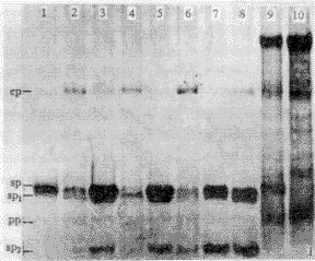
, http://www.100md.com
Fig.1 Electrophoretic analysis of purified SP. Lane 1: further purified SP; lane 2-3: pellet and SP after centrifugation at 112000 g for 180 min; lane 4-5: pellet and SP after 112000 g for 100 min; lane 6-7: pellet and SP after 142 000 g , 180 min; lane 8: SP after 63000 g, 100 min; lane 9: infected sap; lane 10: healthy sap. SP1 and 2: the proteins degraded from SP; PP: plant protein; CP: capsid protein.
图1 提纯SP的电泳分析。1:精提纯的SP:2―3:112 000g离心180min后的沉淀和SP;4―5:112 000g, 100min后的沉淀和SP; 6―7: 142000g,180min后的沉淀和SP;8: 63000g, 100min后的SP;9:病汁液;10:健汁液。SP1和SP2:SP降解产物;PP:植物蛋白;CP:衣壳蛋白。
, 百拇医药
The crude and further purified SP all had a typical ultraviolet absorption spectrum. Here, we showed the spectrum of further purified protein (Fig.2), which had a maximum absorbance at 276 nm, and a minimum absorbance at 255 nm. The 260/280 ratio was about 0.8. Using both methods, a large quantity of SP were produced. The yield obtained by method I was about 2.0 mg/g tissue, and that of method II was about 0.8 mg/g tissue. However, the SP prepared from the latter was purer than that from the former by analysis of ultraviolet absorption (data not shown). Additionally, the method of electro-elution was proved to be efficient to separate protein from polyacrylamide gel with a recovery efficiency of about 80%.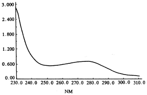
, 百拇医药
Fig.2 Ultraviolet absorption spectrum of further purified SP
图2 精提纯SP的紫外吸收曲线
2 SDS-PAGE analysis of purified SP
After electrophoresis, the SP directly extracted from the infected leaves sap appeared as a single band in the gel with molecular weight of 20.1 kDa. However, the purified SP showed as a major band and a minor band with the size of 20.1 kDa and 18.9 kDa. No corresponding band appeared in the lane of healthy leaves sap (Fig.3), which indicated that the purified protein was exactly the disease-specific protein. It seemed that SP is readily degraded, as shown in figure 1. The minor band must be the smaller peptide degraded from the major band during purification.
, 百拇医药
Fig.3 Electrophoretic analysis of SP
Lane 1,2,3: infected sap; lane 4: marker;
lane 5: purified SP; lane 6: healthy sap.
图3 提纯SP的电泳分析。1,2,3:病汁液;4:标准分子量;5:提纯的SP;6:健汁液。
3 Serological characteristics
The titer of antiserum raised against crude SP was 51 200, with an optimal working concentration at 5000-10000 dilution. However, the antiserum could nonspecifically react with healthy tissue because of the antigen's containing a certain degree of plant protein, especially reacted at the working concentration of 800 dilution and lower. The antiserum against the further purified SP was highly specific with a titer of 6 400. The optimal working concentration was 500-1000. With the highly specific antiserum, we investigated the serological relationship. The results showed that RSV-SP antiserum had weakly reaction with RGSV-SP, and no serological reaction with RSV-CP and RGSV-CP. However, no positive reactions were detected between RSV-SP and the antisera of RGSV-SP, RGSV-CP and RSV-CP (data not shown). These results confirmed that there is evolutionary relationship between RSV and RGSV.
, 百拇医药
3 Discussion
The method for purification of disease-specific protein of RSV according to the principle of precipitation of protein by saturated ammonium sulfate has already been reported by Kiso et al (1973)[14]. However, the procedures were much complicated and the effect was not ideal in practice. Here, we established two methods mainly according to the principle that protein dissolves in buffer of pH7.0 and crystallizes in the buffer of low pH. The crude preparation contained no CP impurity and only a bit of plant protein whose molecular weight is close to that of SP. However, we do not precisely understand why the yield of method II was much lower but purer than that of method I. One reason may be that a large quantity of non-crystallized and indissoluble SP lost during centrifugation, the evidence was that much SP was found in the pellet (Fig.1). In the four combination of centrifuge speed and time, centrifugation at 42000 g for 180 min showed best effect to discard virus particles.However, the Mr of SP is only about 20.1 kDa, it is difficult to non-polluted by a kind of plant protein of 14 kDa. The further purified methods, separating proteins by polyacrylamide gel electrophoresis and recovering by differential pH method and electro-elution, were successful in discarding all the impurities.
, http://www.100md.com
The amounts of SP in the leaves were reported to be proportional to the degree of severity of symptom and the resistance of rice varieties[3,13], which was confirmed by our experiments[14]. SP also had a readily degraded nature[3]. Therefore, for successfully purification and more yield achievement, three factors should be paid attention to: (Ⅰ) The plant tissue used for purification should be fresh leaves with typical stripe symptoms of susceptible varieties; (Ⅱ) All the procedures should be performed at low temperature; (Ⅲ) The time taken for resuspension and crystallization should be sufficient.
, http://www.100md.com
The titer of the antiserum raised against the crude SP was very high, even though it could nonspecifically react with healthy plant sap, thus the new antiserum can be used for diagnosis, identification and epidemiological forecast of rice stripe disease. In comparison with the antiserum against nucleoprotein, this new way has three advantages: (Ⅰ) A large quantity of antigen were very easy to prepare, the yield of SP was 0.8-2.0 mg/g tissue, and that of RSV nucleoprotein was only roughly 10 mg/kg tissue; (Ⅱ) The procedures for purification of SP is rather simple while that of RSV nucleoprotein is complicated and time-consuming; (Ⅲ) The expenditure for purification of SP is much lower than that of RSV nucleoprotein.
, http://www.100md.com
The antiserum of the further purified SP in our experiment owned a low titer, but a high specificity, which can be applied for serological relationship investigation and molecular biological researches. No serological relationship was detected between RSV-SP and the antisera of RGSV-SP, RSV-CP and RGSV-SP. However, the antiserum of RSV-SP could weakly react with RGSV-SP, which was identical to the result of Miranda et al (1995)[11]. This result confirmed that RGSV and RSV are evolutionarily related and SP and CP maybe originated from completely different ancestors. Analysis of amino acid composition also showed that the RSV nucleoprotein and SP were distinct[15]. There was also no serological reaction between MStV CP antiserum and MStV SP or MStV-SP antiserum and MStV-CP either[4]. However, Toriyama (1986) reported that antiserum raised against SP reacted moderately with RSV-CP in the ring interface test,although RSV-CP antiserum did not react with SP[3]. As mentioned above, the RSV-SP used for preparation of antiserum was purified by saturated ammonium sulfate method and contained much impurities including virus particles and plant proteins. So the antiserum was also not pure, together with the plant proteins including in the purified virus preparation, we suggested that the reaction between SP and CP was pseudo-positive reaction.
, 百拇医药
RSV-SP has serological relationship with RGSV-SP even though the amino acid identity between their sequences is only 28.0%. The identities between MStV-SP and RSV-SP, RHBV-SP at amino acid level is 76.8% and 56.9%, respectively[16].However, no serological reaction were investigated between MStV-SP and RSV-SP, RHBV-SP[4,7]. So whether there is serological reaction between two proteins not only depend on sequence identity at amino acid level, but also may be related to secondary conformation of proteins.
, http://www.100md.com
Analysis of complete nucleotide sequence of RSV genome indicates that SP is encoded by viral-sense RNA4 segment[9,10]. However, the function of this protein is unknown. When the deduced SP sequence was compared with the sequences in the NBRF protein data bank, no significant homologies were detected. A hydropathy plot of the SP sequence did not reveal any cluster of changed amino acids or any significant hydrophobic region typical to transmembrane proteins[17]. The evidence that the non-viruliferous insect vectors could transmit virus after injection of purified virus into the abdomen[4,18] showed that SP of Tenuivirus is not necessary for infectivity. Recently, Shimizu et al[19] reported that there is a nonviral sequence in the 5′-termini of mRNAs of RSV-RNA3 and RNA4. Combining with the facts that the content of SP was in proportion to the degree of severity of symptom and the resistance of rice varieties, we suggested that the expression of SP is regulated and controlled by host genes, and SP may be one kind of pathogenetic proteins which disturb the function of chloroplast by unknown mechanisms. Additionally, SP gene of Tenuivirus was reported to share 27%-35% nucleotide identity with aphid-transmitted helper gene found in cauliflower mosaic virus, which suggested that SP may be also involved in vector transmission[21]. Recently, we found that SP was localized in chloroplast by immunogold technique and that two RSV isolates with 100% sequence identity of CP gene and 99% sequence identity of SP gene at amino acid level caused significant different infected occurrence on rice varieties, which suggested that SP was related to cholorotic symptoms and vector recognition (unpublished data). However, to accurately identify the functions of SP, a fully infective cDNA clone of RSV must be constructed. Rice protoplast and insect cell infective systems and a series of delete mutants of SP gene are also required.
, 百拇医药
Acknowledgements This work was supported by the National Natural Science Foundation (item number 39670489) and Natural Science Foundation of Fujian Province (item number C97031).
References
1 Lin Qiying, Xie Lianhui, Zhou Zhongju et al. Studies on rice stripe diseaseI. distribution and loss. Journal of Fujian Agricultural College, 1990,19(4):373~379 (in Chinese)
2 Murphy FA, Fanaquet CM, Bishop DHL et al. Classification and nomenclature of viruses: Sixth Report of the International Committee on Taxonomy of Viruses. Arch Virol, 1995,(suppl. 10):316
, http://www.100md.com
3 Toriyama S. Rice stripe virus: prototype of a new group of viruses that replicated in plants and insects. Microbiol Sci, 1986,(3):347~351
4 Gingery RE, Nault LR, Bradfute OE. Maize stripe virus: characteristics of a member of a new viurs class. Virology, 1981,112:99~108
5 Falk BW, Tsai JH. Assay for maize stripe virus-infected plants by using antiserum produced to a purified noncapsid protein. Phytopathology, 1983,73:1259~1262
, 百拇医药
6 Morales FJ, Nissen AI. Association of spiral filamentous virus-like particles with rice hoja blanca. Phytopathology, 1983,73:971~974
7 Falk BW, Morales FJ, Tsai JH et al. Serological and biochemical properties of the capsid and noncapsid proteins of maize stripe, rice hoja blanca, and Echinochloa hoja blanca virus. Phytopathology, 1987,77:196~201
8 Atabekov JG, Popova GA, Kiselv NA et al. In vitro polymerization of winter wheat mosaic virus antigen. Virology, 1968,35:458~472
, 百拇医药
9 Kakutani T, Hayano Y, Hayashi T et al. Ambisense segment 4 of rice stripe virus: possible evolutionary relationship with phleboviruses and uukuviruses (Bunyaviriade). J Gen Virol, 1990, 71:1427~1432
10 Zhu Y, Hayakawa T, Toriyama S. Complete nucleotide sequence of RNA4 of rice stripe virus isolate T, and comparison with another isolate and with maize stripe virus. J Gen Virol, 1992,73:1309~1312
11 Miranda GJ, Koganezawa H. Identification, purification and serology detection of major noncapsid protein of rice grassy stunt virus. Phytopathology, 1995,85:1530~1533
, http://www.100md.com
12 Lu Jiajue, Zhang Chenglian, Zhang Zuofang. Studies on detection of plant viruses by using protein A-ELISA. Plant Quarantine, 1990,4(5):161~163 (in Chinese)
13 Kiso A, Yamamoto T. Infection and symptom development in rice stripe disease with special reference to disease-specific protein other than virus. Review of Plant Protection Research, 1973, 6:75~100
14 Lin Qitian, Lin Hanxin, Wu Zujian et al. Accumulations of coat protein and disease-specific protein of rice stripe virus in its host. Journal of Fujian Agricultural University, 1998,27(3): 322~326
, 百拇医药
15 Toriyama S. Chemical composition of coat protein of rice stripe virus and stripe disease specific protein. Ann Phytopathol Soc Jpn, 1983,49:432
16 Toriyama S, Kimishima T, Takahashi M. The proteins encoded by rice grassy stunt virus RNA5 and RNA6 are distantly related to the corresponding proteins of other members of the genus Tenuivirus. J Gen Virol, 1997, 78:2355~2363
17 Huiet L, Klaassen V, Tsai JH et al. Identification and sequence analysis of the maize stripe virus major noncapsid protein gene. Virology, 1990, 179:862~866
, http://www.100md.com
18 Toriyama S. Characterizatoin of rice stripe virus: a heavy component carrying infectivity. J Gen Virol, 1982,61:187~195
19 Shimizu T, Toriyama S, Takahashi M et al. Non-viral sequences at the 5′termini of mRNAs derived from virus-sense and virus-complementary sequences of the ambisense RNA segments of rice stripe tenuiviruse. J Gen Virol, 1996,77:541~546
20 Ramirez B-C, Lozano I, Constantino LM et al. Complete nucleotide sequence and coding strategy of rice hoja blanca virus. J Gen Virol, 1993,74:2463~2468, http://www.100md.com