超声测量颈动脉内膜中层厚度与颈动脉斑块的关系
作者:许竹梅 赵水平 范平
单位:许竹梅(湖南医科大学附属第二医院 心内科);赵水平(湖南医科大学附属第二医院 心内科);范平(湖南医科大学附属第二医院 超声室; 湖南省长沙市 410011)
关键词:超声;颈动脉;内膜中层厚度;动脉粥样硬化
中国动脉硬化杂志000222[摘 要] 为了探讨颈动脉内膜中层厚度与局限性颈动脉斑块的联系,对91名受试对象的颈总动脉内膜中层厚度及颈内动脉和颈动脉分叉处的斑块进行超声检测,并将颈动脉内膜中层厚度进行分级。结果观察到有斑块者较无斑块者其颈动脉内膜中层厚度明显增加(0.83±0.16 mm比0.64±0.12 mm, P<0.01),且随斑块的严重程度增加,其内膜中层厚度呈增厚趋势。该结果支持颈总动脉内膜中层厚度与颈动脉局限性动脉粥样硬化斑块明显相关,提示颈动脉内膜中层厚度增厚可能是颈动脉粥样硬化的早期表现。
, 百拇医药
[中图分类号] R543.4 [文献标识码] A
[文章编号] 1007-3949(2000)-02-0165-04
Relation of Itima-Media Thickness to Plaque in Carotid Arteries Measured by Ultrasound
XU Zhu-Mei, ZHAO Shui-Ping, and Fan-Ping
(Department of Cardiology, the Second Affiliated Hospital, Hunan Medical University, Changsha 410011, China)
ABSTRACT Aim The paper was done to evaluate the relations between intima-media thickness (IMT) of common carotid artery (CCA) and plaque in the internal carotid artery (ICA) and bifurcations (CBs). Methods B-mode ultrasound examination of carotid arteries was per formed in 91 subjects. B-mode ultrasound examination include measurement of CCA-IMT and the sites of plaque in the ICA and CBs. Results The results showed that the prevalence of plaque increased from the lowest to the highest quintile of IMT(linear trend: P<0.01). The mean IMT was lower in subjects without plaques than in subjects with plaques (0.64±0.12 mm vs 0.83±0.16 mm,P<0.01), there was an increasing trend for IMT with the severity of plaque. Conclusion Increases in IMT as measured in the CCA are clearly related to atherosclerotic plaque in the ICA and CBs.
, http://www.100md.com
MeSH Ultrasound; Carotid Artery; Intima-Media Thickness; Atherosclerosis
超声能清晰显示体表大动脉特别是颈动脉血管壁的情况,近年来逐渐被用于作为检测早期动脉粥样硬化的无创方法[1]。Pignoli等[2]首先报道了超声测量颈动脉内膜中层厚度(intima-media thickness, IMT)的方法,证实超声能准确测定颈动脉IMT,并认为颈动脉IMT的早期改变会导致以后颈动脉粥样硬化斑块的发生。然而,超声检测颈动脉IMT和局限性颈动脉粥样硬化斑块间的联系较少报道,本文就此进行探讨。
1 资料和方法
1.1 研究对象
91名研究对象,年龄40~74岁,平均年龄55.9±7.3岁,其中男67例,女24例。其中冠心病患者及冠心病高危者65例,正常人26例。
, 百拇医药
1.2 超声检查
彩色超声仪acuson128XP/10, 备有7.0 MHz探头,其轴分辨率为0.1 mm。 B超检查及读数均由我院超声室同一名医师完成。被检查者取仰卧位,颈后薄枕,放松,头部偏向检查区对侧,先从锁骨内侧端横向检查颈总动脉,然后将探头沿其走行方向向头部移动,逐节段从前、侧、后三个方向观察双侧颈总动脉(common carotid artery, CCA)、颈内动脉(internal carotid artery, ICA)和颈动脉分叉(carotid bifurcation, CB)的横轴及纵轴实时二维图像,并尽可能检查至颈部最高部位。
1.2.1 颈总动脉内膜中层厚度测量 横纵向观察颈总动脉,颈总动脉后壁纵向超声显像可表现为由相对较低回声分隔的两条平行亮线,即“双线型 ”图像。内线为内膜与管腔的分界线,外线为中外膜分界线,其间距离即为IMT,见图1(Figure 1)。冻结颈动脉窦以下1 cm处(无斑块部位)的图像,共测量5次IMT,取10次左右侧测量值的平均值即为平均颈总动脉IMT。在测量颈总动脉IMT的部位时同时测量其颈总动脉内径,取10次平均值。每次均在同步心电图记录的R波顶端冻结图像。本组91名受试者的颈总动脉IMT均可被高分辨率B超清晰检测,测定的IMT范围在0.44~1.20 mm间。根据颈总动脉IMT,将受试者分为三组,即IMT≤0.60 mm组、IMT 0.61~0.79 mm组和IMT≥0.80 mm组。
, 百拇医药
图1. 颈动脉内膜中层厚度示意图.
Figure 1. Schematic diagram for measuring carotid arterial intima-media thickness. Line 1, represents interface between periadventitia and adventitia of near wall; line 2, adventitia-media interface of near wall; line 3, intima-lumen interface of near wall; line 4, lumen-intima interface of far wall; line 5, media-adventitia interface of far wall; line 6, adventitia-periadventitia interface of far wall. Distance between lines 4 and 5 corresponds the far wall intima-media thickness.
, 百拇医药
1.2.2 斑块测定 斑块定义为局限性回声结构突出管腔(回声可不均匀或伴声影),厚度≥1.3 mm。观察以上三组颈内动脉和颈动脉分叉处斑块情况,若发现斑块,横纵向冻结图像,并测量其厚度,若在同一动脉段发现多个斑块,记录斑块数目,并测量最大斑块厚度。半定量估计斑块的范围及严重程度:0级为无斑块;1级为单侧斑块≤2 mm;2级为单侧斑块>2 mm或双侧均有斑块且其中至少一侧斑块≤2 mm;3级为双侧斑块均>2 mm。
1.3 危险因素调查
对各研究对象进行血压、血脂及年龄各项心血管危险因素的测定。包括收缩压、总胆固醇、甘油三酯、载脂蛋白B100和低密度脂蛋白胆固醇等的测定。
1.4 统计学方法
所有统计学处理均采用SPSS统计软件包进行。计量资料采用均数±标准差,随着IMT增加,斑块发生率的线性趋势采用线性回归,不同颈动脉分级的颈动脉斑块发生率采用卡方检验,颈总动脉IMT与颈内动脉和颈动脉分叉处斑块严重程度的关系采用方差分析。各危险因素与颈动脉IMT的关系采用Spearman’s相关分析,并进行逐步回归分析以判断危险因素对颈动脉IMT的影响。颈总动脉斑块分级间危险因素的比较采用方差分析。
, 百拇医药
2 结 果
高分辨率B型超声可清晰显示体表大动脉血管壁情况,本文中所有受试者颈动脉IMT都清晰可辨,B超能清楚显示颈动脉内的粥样斑块,提示该法可作为检测动脉粥样硬化的无创方法(图2和3,Figure 2 and 3)。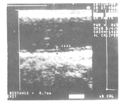
图2. 正常颈动脉内膜中层厚度超声图.
Figure 2. B-mode sonogram of normal carotid arterial intima-media thickness . Distance between the arrowheads corresponds to far wall intima-media thickness. CCA, common carotid artery.
, 百拇医药 内膜中层厚度≤0.60 mm组、内膜中层厚度为0.61~0.79 mm组和内膜中层厚度≥0.80 mm组粥样硬化斑块发生率分别为18.5%、37.5%和75 %(表1,Table 1),三组之间有明显统计学差异(P<0.01)。经线性回归分析,发现颈总动脉IMT与颈内动脉和颈动脉分叉处斑块的发生率呈正相关(r=0.529,P<0.01,Y=0.126+0.697X)。
图3. 颈动脉斑块的超声图.
Figure 3. B-mode sonogram of carotid arterial plaque. Abnormal artery with focal protrusion of wall into lumen (arrow), the thickness is more than 1.3 mm,representing carotid plaque.
, 百拇医药
表1. 不同颈总动脉内膜中层厚度分级与颈内动脉和颈动脉分叉处斑块分级情况.
Table 1. Prevalence of plaque of the CB-ICA according to quintiles of CCA-IMT (mm). Plaque
≤0.6
n(%)
0.61~0.79
n(%)
≥0.80
n(%)
0
22 (81.5)
, 百拇医药
20 (62.5)b
8 (25.0)ab
1
3 (11.1)
6 (18.8)b
7 (21.9)ab
2
2 (7.4)
4 (12.5)b
8 (25.0)ab
, http://www.100md.com 3
0 (0)
2 (6.2)b
9 (28.1)ab
Total
27 (100.0)
32(100.0)b
32 (100)ab
a: P<0.01,compared with IMT 0.61~0.79 mm group, b: P<0.01,compared with IMT ≤0.61 mm group.
, 百拇医药
根据受试者颈动脉内是否检测到粥样斑块进行分组,观察到有粥样斑块组IMT平均值(0.83±0.16 mm)明显高于无粥样斑块组(0.64±0.12 mm),两组间有显著差别(P<0.01)。进一步研究发现随斑块的严重程度增加,颈总动脉IMT呈增厚趋势(表2,Table 2)。
血压、血脂和年龄等与颈动脉内膜中层厚度有明显关系,Spearman’s相关分析发现年龄(r=0.296,P<0.05)、收缩压(r=0.348,P<0.05)、总胆固醇(r=0.348,P<0.05)、甘油三酯(r=0.28,P<0.01)、低密度脂蛋白胆固醇(r=0.315,P<0.05)和载脂蛋白B100(r=316,P<0.05)等心血管危险因素与颈动脉IMT呈明显正相关,逐步回归分析发现收缩压、总胆固醇、年龄、甘油三酯和载脂蛋白B100对颈动脉IMT具有显著回归效果。回归方程为Y=-0.392+0.003028×收缩压+0.02593×总胆固醇+0.006898×年龄+0.03214×甘油三酯+0.105×B100(表3,Table 3)。
, 百拇医药
表2. 斑块严重程度与颈动脉内膜中层厚度的关系.
Table 2. Relation between CCA-IMT and plaques of ICA and CB. Plaque
n
IMT(mm)
No plaque (0)
50
0.64±0.12
Mild plaque(1)
16
0.81±0.17a
, 百拇医药
Modest plaque(2)
14
0.82±0.16a
Severe plaque(3)
11
0.89±0.11b
a: P<0.05,compared with no plaque group; b:P<0.05,compared with no, mild and modest plaque group.表3. 颈动脉内膜中层厚度分级与部分心血管危险因素.
Table 3. Associations between CCA-IMT with some cardiovascular risk factors( ±s). IMT
, 百拇医药
≤0.60 mm
0.61~0.79 mm
≥0.80 mm
n
27
32
32
IMT(mm)
0.54±0.05
0.70±0.05
0.92±0.09
LD (mm)
, http://www.100md.com
6.21±0.37
6.64±0.79
6.56±0.87
Clinical data
Age(years)
53±6
57±8
58±8a
SBP(mmHg)
122±14
141±22
145±23a
, 百拇医药
TC (mmol/L)
5.02±0.83
5.35±1.02
6.13±1.58b
LDLC(mmol/L)
2.92±0.75
3.14±0.88
3.68±0.07b
HDLC(mmol/L)
1.40±0.30
1.36±0.66
, 百拇医药
1.35±0.32c
TG(mmol/L)
1.49±0.69
1.99±1.98
2.42±2.13b
BS(mmol/L)
4.68±0.60
5.27±0.61
5.22±0.86c
ApoB100(g/L)
0.96±0.18
, http://www.100md.com
1.10±0.30
1.21±0.30b
ApoAI(g/L)
1.33±0.32
1.25±0.24
1.26±0.22c
a: P<0.01, b: P<0.05, c: NS.
3 讨 论
超声检测的动脉粥样硬化主要表现为以下两种类型:血管壁增厚和粥样斑块。B超发现动脉粥样斑块能直观证明动脉粥样硬化病变的存在,但并不是所有动脉粥样硬化者均能检测到粥样斑块,因为在动脉粥样硬化的早期,动脉粥样硬化可仅表现为血管壁增厚。近年来许多研究者认为动脉IMT能反映血管壁增厚情况,有人将超声方法与病理组织学方法进行比较,发现超声能准确测量动脉IMT ,且动脉粥样硬化时IMT的改变早于斑块的发生[2,3]。我们研究表明动脉粥样硬化的危险因素(如年龄、脂质代谢紊乱、吸烟、高血压等) 与颈动脉IMT增厚相关,与文献[4,5]报告一致。家族性高胆固醇血症患者在儿童期就出现颈动脉IMT增厚[6,7],O’leary 等[8]发现颈动脉IMT是心肌梗死和脑卒中的危险因素,这些研究结果支持超声测量到颈动脉IMT增厚是一种早期反映动脉粥样硬化的无创性指标。最近Raitakari 等[9]将颈动脉内膜中层厚度和血管内皮功能作为早期动脉粥样硬化的指标,研究脂蛋白(a)导致动脉粥样硬化的机制。
, 百拇医药
颈总动脉形态直,位置表浅,且与皮肤平行,故颈总动脉IMT容易测量,且颈总动脉IMT测量值可靠,重复性好。所以,我们把颈总动脉作为测量IMT的部位。此外,B超定义的IMT增厚有其模糊性,可以指动脉壁的弥漫性增厚如测量平均颈动脉IMT,也可指偏心的增厚或是已确定的动脉粥样硬化增厚(如最大IMT和斑块部位的IMT)。本研究中IMT测量是在颈动脉窦下1 cm无斑块部位,取测量的平均值而非最大值,这样就避开了不规则及弯曲的颈动脉窦及分叉,避免了由于血流动力学紊乱和血管张力改变导致的生理性血管增厚,因此本研究中IMT增厚代表了弥漫的血管壁增厚而非偏心性增厚和局限性动脉粥样斑块。尽管少数研究者不认为这种弥漫性血管壁增厚是动脉粥样硬化的改变[10],但我们发现颈总动脉平均IMT不仅与颈内动脉及颈动脉分叉部斑块的存在有关,且与斑块的严重程度密切相关。随斑块的严重程度增加,颈总动脉IMT呈增厚趋势,支持颈动脉IMT增厚是动脉粥样硬化早期的改变。
有研究表明作为动态定量观察早期动脉粥样硬化进展或消退的指标,颈动脉IMT较斑块可能更有价值。Wendelhag等[11]对降脂治疗2年及5年前后颈动脉斑块面积和IMT的动态变化进行研究,发现尽管两者的变化呈正相关,但追踪观察发现仅21%的研究对象能获定量的斑块测量,而大部分研究对象能获得IMT的定量测定值。所以,颈动脉IMT测定值得在临床上推广应用。
, 百拇医药
[作者简介] 许竹梅,女,1967年生,主治医师。 赵水平,男,1954年生,博士研究生导师。
参考文献
[1] Salonen JT, Salonen R. Ultrasound B-mode in observational studies of atherosclerotic progress [J]. Circulation, 1993, 88: 20-28
[2] Pignoli P, Tremoli E, Poli A, et al. Intimal plus medial thickness of the arterial wall: A direct measurement with ultrasound imaging [J]. Circulation, 1986, 74: 1 399-406
, 百拇医药 [3] Wong M,Edelstein J, Wollman J,et al. Ultrasonic pathological comparison of human arterial wall: Verification of intima-media thickness [J]. Arterioscler Thromb, 1993, 13: 482-486
[4] Gnasso A, Irace C, Pujia PLM. Carotid intima-media thickness and coronary heart risk facts [J]. Atherosclerosis, 1996, 119: 7-15
[5] Skoglund C, Tang R, Bond MG, et al. LDL particle size distribution is associated with carotid intima-media thickness in healthy 50-year-old men [J]. Arterioscler Thromb Vasc Biol, 1999, 19: 2 422-430
, http://www.100md.com
[6] Tonstand S, Joakimsen O, Stensland-Bugge E, et al. Risk facts related to carotid intima-media thickness and plaque in children with familiar hypercholesterolemia and control subjects [J]. Arterioscler Thromb, 1996, 16: 984-991
[7] Pauciullo P, Iannuzzi A, Sartorio R, et al. Increased intima-media thickness of common carotid artery in hypercholesterolemic children [J]. Arterioscler Thromb, 1994, 14: 1 075-079
[8] O'leary DH, Polak JF, Kronmal RA, et al. Carotid artery intima and media thickness as a risk factor for myocardial infarction and stroke in older adults [J]. N Engl J Med, 1999, 340: 14-22
, http://www.100md.com
[9] Raitakari OT, Adams MR, Celermajer DS. Effect of lipoprotein(a) on the early functional and structural changes of atherosclerosis [J]. Arterioscler Thromb Vasc Biol, 1999, 19: 990-995
[10] Zureik M, Touboul PJ, Bonithon-Kopp C, et al. Differential association of common carotid intima-media thickness and carotid atherosclerotic plaques with parental history of premature death from coronary heart disease [J]. Arterioscler Thromb Vasc Biol, 1999, 19: 366-371
, http://www.100md.com
[11] Wendelhag I, Wiklund O, Wkstand J. On quantifying plaque size and intima media thickness in carotid and femoral arteries: Comments on result from a prospective ultrasound study in patients with familiar hypercholesterolemia [J]. Arterioscler Thromb, 1996, 16: 843-850
1999-07-12 收到
2000-02-25 修回, 百拇医药
单位:许竹梅(湖南医科大学附属第二医院 心内科);赵水平(湖南医科大学附属第二医院 心内科);范平(湖南医科大学附属第二医院 超声室; 湖南省长沙市 410011)
关键词:超声;颈动脉;内膜中层厚度;动脉粥样硬化
中国动脉硬化杂志000222[摘 要] 为了探讨颈动脉内膜中层厚度与局限性颈动脉斑块的联系,对91名受试对象的颈总动脉内膜中层厚度及颈内动脉和颈动脉分叉处的斑块进行超声检测,并将颈动脉内膜中层厚度进行分级。结果观察到有斑块者较无斑块者其颈动脉内膜中层厚度明显增加(0.83±0.16 mm比0.64±0.12 mm, P<0.01),且随斑块的严重程度增加,其内膜中层厚度呈增厚趋势。该结果支持颈总动脉内膜中层厚度与颈动脉局限性动脉粥样硬化斑块明显相关,提示颈动脉内膜中层厚度增厚可能是颈动脉粥样硬化的早期表现。
, 百拇医药
[中图分类号] R543.4 [文献标识码] A
[文章编号] 1007-3949(2000)-02-0165-04
Relation of Itima-Media Thickness to Plaque in Carotid Arteries Measured by Ultrasound
XU Zhu-Mei, ZHAO Shui-Ping, and Fan-Ping
(Department of Cardiology, the Second Affiliated Hospital, Hunan Medical University, Changsha 410011, China)
ABSTRACT Aim The paper was done to evaluate the relations between intima-media thickness (IMT) of common carotid artery (CCA) and plaque in the internal carotid artery (ICA) and bifurcations (CBs). Methods B-mode ultrasound examination of carotid arteries was per formed in 91 subjects. B-mode ultrasound examination include measurement of CCA-IMT and the sites of plaque in the ICA and CBs. Results The results showed that the prevalence of plaque increased from the lowest to the highest quintile of IMT(linear trend: P<0.01). The mean IMT was lower in subjects without plaques than in subjects with plaques (0.64±0.12 mm vs 0.83±0.16 mm,P<0.01), there was an increasing trend for IMT with the severity of plaque. Conclusion Increases in IMT as measured in the CCA are clearly related to atherosclerotic plaque in the ICA and CBs.
, http://www.100md.com
MeSH Ultrasound; Carotid Artery; Intima-Media Thickness; Atherosclerosis
超声能清晰显示体表大动脉特别是颈动脉血管壁的情况,近年来逐渐被用于作为检测早期动脉粥样硬化的无创方法[1]。Pignoli等[2]首先报道了超声测量颈动脉内膜中层厚度(intima-media thickness, IMT)的方法,证实超声能准确测定颈动脉IMT,并认为颈动脉IMT的早期改变会导致以后颈动脉粥样硬化斑块的发生。然而,超声检测颈动脉IMT和局限性颈动脉粥样硬化斑块间的联系较少报道,本文就此进行探讨。
1 资料和方法
1.1 研究对象
91名研究对象,年龄40~74岁,平均年龄55.9±7.3岁,其中男67例,女24例。其中冠心病患者及冠心病高危者65例,正常人26例。
, 百拇医药
1.2 超声检查
彩色超声仪acuson128XP/10, 备有7.0 MHz探头,其轴分辨率为0.1 mm。 B超检查及读数均由我院超声室同一名医师完成。被检查者取仰卧位,颈后薄枕,放松,头部偏向检查区对侧,先从锁骨内侧端横向检查颈总动脉,然后将探头沿其走行方向向头部移动,逐节段从前、侧、后三个方向观察双侧颈总动脉(common carotid artery, CCA)、颈内动脉(internal carotid artery, ICA)和颈动脉分叉(carotid bifurcation, CB)的横轴及纵轴实时二维图像,并尽可能检查至颈部最高部位。
1.2.1 颈总动脉内膜中层厚度测量 横纵向观察颈总动脉,颈总动脉后壁纵向超声显像可表现为由相对较低回声分隔的两条平行亮线,即“双线型 ”图像。内线为内膜与管腔的分界线,外线为中外膜分界线,其间距离即为IMT,见图1(Figure 1)。冻结颈动脉窦以下1 cm处(无斑块部位)的图像,共测量5次IMT,取10次左右侧测量值的平均值即为平均颈总动脉IMT。在测量颈总动脉IMT的部位时同时测量其颈总动脉内径,取10次平均值。每次均在同步心电图记录的R波顶端冻结图像。本组91名受试者的颈总动脉IMT均可被高分辨率B超清晰检测,测定的IMT范围在0.44~1.20 mm间。根据颈总动脉IMT,将受试者分为三组,即IMT≤0.60 mm组、IMT 0.61~0.79 mm组和IMT≥0.80 mm组。
, 百拇医药
图1. 颈动脉内膜中层厚度示意图.
Figure 1. Schematic diagram for measuring carotid arterial intima-media thickness. Line 1, represents interface between periadventitia and adventitia of near wall; line 2, adventitia-media interface of near wall; line 3, intima-lumen interface of near wall; line 4, lumen-intima interface of far wall; line 5, media-adventitia interface of far wall; line 6, adventitia-periadventitia interface of far wall. Distance between lines 4 and 5 corresponds the far wall intima-media thickness.
, 百拇医药
1.2.2 斑块测定 斑块定义为局限性回声结构突出管腔(回声可不均匀或伴声影),厚度≥1.3 mm。观察以上三组颈内动脉和颈动脉分叉处斑块情况,若发现斑块,横纵向冻结图像,并测量其厚度,若在同一动脉段发现多个斑块,记录斑块数目,并测量最大斑块厚度。半定量估计斑块的范围及严重程度:0级为无斑块;1级为单侧斑块≤2 mm;2级为单侧斑块>2 mm或双侧均有斑块且其中至少一侧斑块≤2 mm;3级为双侧斑块均>2 mm。
1.3 危险因素调查
对各研究对象进行血压、血脂及年龄各项心血管危险因素的测定。包括收缩压、总胆固醇、甘油三酯、载脂蛋白B100和低密度脂蛋白胆固醇等的测定。
1.4 统计学方法
所有统计学处理均采用SPSS统计软件包进行。计量资料采用均数±标准差,随着IMT增加,斑块发生率的线性趋势采用线性回归,不同颈动脉分级的颈动脉斑块发生率采用卡方检验,颈总动脉IMT与颈内动脉和颈动脉分叉处斑块严重程度的关系采用方差分析。各危险因素与颈动脉IMT的关系采用Spearman’s相关分析,并进行逐步回归分析以判断危险因素对颈动脉IMT的影响。颈总动脉斑块分级间危险因素的比较采用方差分析。
, 百拇医药
2 结 果
高分辨率B型超声可清晰显示体表大动脉血管壁情况,本文中所有受试者颈动脉IMT都清晰可辨,B超能清楚显示颈动脉内的粥样斑块,提示该法可作为检测动脉粥样硬化的无创方法(图2和3,Figure 2 and 3)。
图2. 正常颈动脉内膜中层厚度超声图.
Figure 2. B-mode sonogram of normal carotid arterial intima-media thickness . Distance between the arrowheads corresponds to far wall intima-media thickness. CCA, common carotid artery.
, 百拇医药 内膜中层厚度≤0.60 mm组、内膜中层厚度为0.61~0.79 mm组和内膜中层厚度≥0.80 mm组粥样硬化斑块发生率分别为18.5%、37.5%和75 %(表1,Table 1),三组之间有明显统计学差异(P<0.01)。经线性回归分析,发现颈总动脉IMT与颈内动脉和颈动脉分叉处斑块的发生率呈正相关(r=0.529,P<0.01,Y=0.126+0.697X)。
图3. 颈动脉斑块的超声图.
Figure 3. B-mode sonogram of carotid arterial plaque. Abnormal artery with focal protrusion of wall into lumen (arrow), the thickness is more than 1.3 mm,representing carotid plaque.
, 百拇医药
表1. 不同颈总动脉内膜中层厚度分级与颈内动脉和颈动脉分叉处斑块分级情况.
Table 1. Prevalence of plaque of the CB-ICA according to quintiles of CCA-IMT (mm). Plaque
≤0.6
n(%)
0.61~0.79
n(%)
≥0.80
n(%)
0
22 (81.5)
, 百拇医药
20 (62.5)b
8 (25.0)ab
1
3 (11.1)
6 (18.8)b
7 (21.9)ab
2
2 (7.4)
4 (12.5)b
8 (25.0)ab
, http://www.100md.com 3
0 (0)
2 (6.2)b
9 (28.1)ab
Total
27 (100.0)
32(100.0)b
32 (100)ab
a: P<0.01,compared with IMT 0.61~0.79 mm group, b: P<0.01,compared with IMT ≤0.61 mm group.
, 百拇医药
根据受试者颈动脉内是否检测到粥样斑块进行分组,观察到有粥样斑块组IMT平均值(0.83±0.16 mm)明显高于无粥样斑块组(0.64±0.12 mm),两组间有显著差别(P<0.01)。进一步研究发现随斑块的严重程度增加,颈总动脉IMT呈增厚趋势(表2,Table 2)。
血压、血脂和年龄等与颈动脉内膜中层厚度有明显关系,Spearman’s相关分析发现年龄(r=0.296,P<0.05)、收缩压(r=0.348,P<0.05)、总胆固醇(r=0.348,P<0.05)、甘油三酯(r=0.28,P<0.01)、低密度脂蛋白胆固醇(r=0.315,P<0.05)和载脂蛋白B100(r=316,P<0.05)等心血管危险因素与颈动脉IMT呈明显正相关,逐步回归分析发现收缩压、总胆固醇、年龄、甘油三酯和载脂蛋白B100对颈动脉IMT具有显著回归效果。回归方程为Y=-0.392+0.003028×收缩压+0.02593×总胆固醇+0.006898×年龄+0.03214×甘油三酯+0.105×B100(表3,Table 3)。
, 百拇医药
表2. 斑块严重程度与颈动脉内膜中层厚度的关系.
Table 2. Relation between CCA-IMT and plaques of ICA and CB. Plaque
n
IMT(mm)
No plaque (0)
50
0.64±0.12
Mild plaque(1)
16
0.81±0.17a
, 百拇医药
Modest plaque(2)
14
0.82±0.16a
Severe plaque(3)
11
0.89±0.11b
a: P<0.05,compared with no plaque group; b:P<0.05,compared with no, mild and modest plaque group.表3. 颈动脉内膜中层厚度分级与部分心血管危险因素.
Table 3. Associations between CCA-IMT with some cardiovascular risk factors(
±s). IMT, 百拇医药
≤0.60 mm
0.61~0.79 mm
≥0.80 mm
n
27
32
32
IMT(mm)
0.54±0.05
0.70±0.05
0.92±0.09
LD (mm)
, http://www.100md.com
6.21±0.37
6.64±0.79
6.56±0.87
Clinical data
Age(years)
53±6
57±8
58±8a
SBP(mmHg)
122±14
141±22
145±23a
, 百拇医药
TC (mmol/L)
5.02±0.83
5.35±1.02
6.13±1.58b
LDLC(mmol/L)
2.92±0.75
3.14±0.88
3.68±0.07b
HDLC(mmol/L)
1.40±0.30
1.36±0.66
, 百拇医药
1.35±0.32c
TG(mmol/L)
1.49±0.69
1.99±1.98
2.42±2.13b
BS(mmol/L)
4.68±0.60
5.27±0.61
5.22±0.86c
ApoB100(g/L)
0.96±0.18
, http://www.100md.com
1.10±0.30
1.21±0.30b
ApoAI(g/L)
1.33±0.32
1.25±0.24
1.26±0.22c
a: P<0.01, b: P<0.05, c: NS.
3 讨 论
超声检测的动脉粥样硬化主要表现为以下两种类型:血管壁增厚和粥样斑块。B超发现动脉粥样斑块能直观证明动脉粥样硬化病变的存在,但并不是所有动脉粥样硬化者均能检测到粥样斑块,因为在动脉粥样硬化的早期,动脉粥样硬化可仅表现为血管壁增厚。近年来许多研究者认为动脉IMT能反映血管壁增厚情况,有人将超声方法与病理组织学方法进行比较,发现超声能准确测量动脉IMT ,且动脉粥样硬化时IMT的改变早于斑块的发生[2,3]。我们研究表明动脉粥样硬化的危险因素(如年龄、脂质代谢紊乱、吸烟、高血压等) 与颈动脉IMT增厚相关,与文献[4,5]报告一致。家族性高胆固醇血症患者在儿童期就出现颈动脉IMT增厚[6,7],O’leary 等[8]发现颈动脉IMT是心肌梗死和脑卒中的危险因素,这些研究结果支持超声测量到颈动脉IMT增厚是一种早期反映动脉粥样硬化的无创性指标。最近Raitakari 等[9]将颈动脉内膜中层厚度和血管内皮功能作为早期动脉粥样硬化的指标,研究脂蛋白(a)导致动脉粥样硬化的机制。
, 百拇医药
颈总动脉形态直,位置表浅,且与皮肤平行,故颈总动脉IMT容易测量,且颈总动脉IMT测量值可靠,重复性好。所以,我们把颈总动脉作为测量IMT的部位。此外,B超定义的IMT增厚有其模糊性,可以指动脉壁的弥漫性增厚如测量平均颈动脉IMT,也可指偏心的增厚或是已确定的动脉粥样硬化增厚(如最大IMT和斑块部位的IMT)。本研究中IMT测量是在颈动脉窦下1 cm无斑块部位,取测量的平均值而非最大值,这样就避开了不规则及弯曲的颈动脉窦及分叉,避免了由于血流动力学紊乱和血管张力改变导致的生理性血管增厚,因此本研究中IMT增厚代表了弥漫的血管壁增厚而非偏心性增厚和局限性动脉粥样斑块。尽管少数研究者不认为这种弥漫性血管壁增厚是动脉粥样硬化的改变[10],但我们发现颈总动脉平均IMT不仅与颈内动脉及颈动脉分叉部斑块的存在有关,且与斑块的严重程度密切相关。随斑块的严重程度增加,颈总动脉IMT呈增厚趋势,支持颈动脉IMT增厚是动脉粥样硬化早期的改变。
有研究表明作为动态定量观察早期动脉粥样硬化进展或消退的指标,颈动脉IMT较斑块可能更有价值。Wendelhag等[11]对降脂治疗2年及5年前后颈动脉斑块面积和IMT的动态变化进行研究,发现尽管两者的变化呈正相关,但追踪观察发现仅21%的研究对象能获定量的斑块测量,而大部分研究对象能获得IMT的定量测定值。所以,颈动脉IMT测定值得在临床上推广应用。
, 百拇医药
[作者简介] 许竹梅,女,1967年生,主治医师。 赵水平,男,1954年生,博士研究生导师。
参考文献
[1] Salonen JT, Salonen R. Ultrasound B-mode in observational studies of atherosclerotic progress [J]. Circulation, 1993, 88: 20-28
[2] Pignoli P, Tremoli E, Poli A, et al. Intimal plus medial thickness of the arterial wall: A direct measurement with ultrasound imaging [J]. Circulation, 1986, 74: 1 399-406
, 百拇医药 [3] Wong M,Edelstein J, Wollman J,et al. Ultrasonic pathological comparison of human arterial wall: Verification of intima-media thickness [J]. Arterioscler Thromb, 1993, 13: 482-486
[4] Gnasso A, Irace C, Pujia PLM. Carotid intima-media thickness and coronary heart risk facts [J]. Atherosclerosis, 1996, 119: 7-15
[5] Skoglund C, Tang R, Bond MG, et al. LDL particle size distribution is associated with carotid intima-media thickness in healthy 50-year-old men [J]. Arterioscler Thromb Vasc Biol, 1999, 19: 2 422-430
, http://www.100md.com
[6] Tonstand S, Joakimsen O, Stensland-Bugge E, et al. Risk facts related to carotid intima-media thickness and plaque in children with familiar hypercholesterolemia and control subjects [J]. Arterioscler Thromb, 1996, 16: 984-991
[7] Pauciullo P, Iannuzzi A, Sartorio R, et al. Increased intima-media thickness of common carotid artery in hypercholesterolemic children [J]. Arterioscler Thromb, 1994, 14: 1 075-079
[8] O'leary DH, Polak JF, Kronmal RA, et al. Carotid artery intima and media thickness as a risk factor for myocardial infarction and stroke in older adults [J]. N Engl J Med, 1999, 340: 14-22
, http://www.100md.com
[9] Raitakari OT, Adams MR, Celermajer DS. Effect of lipoprotein(a) on the early functional and structural changes of atherosclerosis [J]. Arterioscler Thromb Vasc Biol, 1999, 19: 990-995
[10] Zureik M, Touboul PJ, Bonithon-Kopp C, et al. Differential association of common carotid intima-media thickness and carotid atherosclerotic plaques with parental history of premature death from coronary heart disease [J]. Arterioscler Thromb Vasc Biol, 1999, 19: 366-371
, http://www.100md.com
[11] Wendelhag I, Wiklund O, Wkstand J. On quantifying plaque size and intima media thickness in carotid and femoral arteries: Comments on result from a prospective ultrasound study in patients with familiar hypercholesterolemia [J]. Arterioscler Thromb, 1996, 16: 843-850
1999-07-12 收到
2000-02-25 修回, 百拇医药