ΗΙ«ΜΉΔ…δLPSΚσ¥σ σΡ‘ΡΎΝΉΥαΜ·p44/42 MAPK±μ¥οΒΡ±δΜ·
Ής’ΏΘΚΆθξΊ ΆθΑΌ»Χ ΜΤΈΡΫζ ΕΈ–Γάρ ΨœΙΣ
ΒΞΈΜΘΚΒΎΥΡΨϋ“Ϋ¥σ―ß…ώΨ≠ΩΤ―ß―–ΨΩΥυ…ώΨ≠Οβ“ΏΒςΫΎ―–ΨΩ “Θ§ΈςΑ≤710032
ΙΊΦϋ¥ ΘΚΝΉΥαΜ·ΒΡp44/42 MAPKΘΜ÷§ΕύΧ«(LPS)ΘΜΡ‘ΘΜ¥σ σ
ΫβΤ ―ß±®00zk13
ΓΨ’Σ“ΣΓΩΡΩΒΡ ―–ΨΩΗΙ«ΜΉΔ…δLPSΚσ¥σ σΡ‘ΡΎΜνΜ·–Έ ΫΒΡœΗΑϊΆβ–≈Κ≈ ΒςΫΎΒΑΑΉΦΛΟΗ(p44/42 MAPK)Ζ÷≤ΦΒΡ±δΜ·ΓΘΖΫΖ®”ΟΟβ“ΏΉι÷·Μ·―ßΖ®ΓΘ ΫαΙϊ ‘ΎΗτΆβ≤ύΚΥΓΔκθœ¬«πΡ‘ΚΥΓΔ ”«Α«χΓΔ–”» ÷–―κΚΥΓΔΈ≤≤ύΚΘ¬μ œ¬«πΡ‘ “≈‘ΚΥΓΔ ”…œΚΥΓΔ»ιΆΖΧε…œΚΥΓΔ÷–―κΜ“÷ ΗΙΆβ≤ύ≤ΩΓΔ±έ≈‘Άβ≤ύΚΥΓΔάΕΑΏΒ»―τ–‘œΗΑϊ ΐΝΩ‘ωΦ” Θ§»Ψ…Ϊ‘ω«ΩΓΘΫα¬έ p44/42 MAPKΒΡΙΠΡή≤ΜΫω”κœΗΑϊΒΡ…ζ≥ΛΓΔΖ÷Μ· ΚΆ…ώΨ≠‘ΣΒΡΩ…Υή–‘±δΜ·”–ΙΊΘ§ΜΙΩ…Ρή”κΟβ“Ώ¥ΧΦΛ”–ΙΊΓΘ
ΓΨ÷–ΆΦΖ÷άύΚ≈ΓΩR322.8 ΓΨΈΡœΉ±ξ Ε¬κΓΩA ΓΨΈΡ’¬±ύΚ≈ΓΩ0529-1356(2000)-‘ω-48
, ΑΌΡ¥“Ϋ“©
THE CHANGES OF THE EXPRESSION OF PHOSPHORYLATED-p44/42 MAPK IN RAT BRAIN AFTER INTRAPERITONEAL INJECTION OF LPS
WANG XiȧWANG Bai-renȧHUANG Wen-jinȧDUAN Xiao-liȧJU Gong*
(Department of NeuroimmunomodulationΘ§Institute of NeuroscienceΘ§the F ourth Medical UniversityΘ§Xi§πan 710032Θ§China)
ΓΨAbstractΓΩObjective To study the changes of the expressio n of phosphorylated-p44/42MAPK in rat brain after intraperitoneal injection of L P S.Method Immunohistochemical staining techniques were used,an d immunostained sections were observed and analyzed under a microscope. Results The expression of phosphorylated-p44/42MAPK increased in la teral septumΘ§septohypothalamic nucleusΘ§preoptic areaΘ§central nucleus of amygd alaΘ§caudal hyppocampusΘ§hypothalamus paraventricular nucleusΘ§supraoptic nucleu sΘ§supramammilary nucleusΘ§ventrolateral part of periaqueductal grayΘ§lateral pa rabranchial nucleusΘ§locus nucleusΘ§et al.Conclusion It sugge s ted that p44/42 MAPK is related with not only cellular growthΘ§differentiation a nd plasticity of neuron but also immune stimuli probably.
, http://www.100md.com
ΓΨkey wordsΓΩp44/42MAPKΘΜ LPSΘΜ BrainΘΜ Rat ΫϋΡξά¥ΒΡ―–ΨΩ±μΟςΘ§÷– ύ…ώΨ≠œΒΆ≥Ω…“‘Ϋ” ήΟβ“Ώ–≈œΔ≤Δ≤Έ”κΕ‘Οβ“ΏΖ¥”ΠΒΡΒςΫΎΘ§ΒΪΕ‘”ΎΡ‘ΡΎ ≤Έ”κΟβ“ΏΖ¥”ΠΒΡ…ώΨ≠ΚΥΆ≈…–≤Μ«ε≥ΰΓΘ‘Ύ…ώΨ≠œΒΆ≥ΙΠΡήΚΆ–ΈΧ§œύΫαΚœΒΡ―–ΨΩ÷–Θ§Φ»Άυ”Π”ΟΫœΕύ ΒΡ «Φ¥ΩΧ‘γΤΎΜυ“ρc-fosΘ§Ά§ ±egr-1Β»“≤”–”Π”ΟΓΘ Υδ »Μ≤ΜΆ§ΒΡ”ΠΦΛΩ…“ΐΤπΡ‘ΡΎ≤ΜΆ§–Έ ΫΒΡΦ¥ΩΧ‘γΤΎΜυ“ρΒΡ±μ¥οΘ§ΒΪ «ΟΩ÷÷Φ¥ΩΧ‘γΤΎΜυ“ρ‘ΎΡ‘ΡΎΒΡ ±μ¥οΨυ”–“ΜΕ®ΒΡΨ÷œό–‘ΓΘ“ρ¥ΥΘ§–η“ΣΫαΚœΕύ÷÷±ξ÷ΨΈο≤≈ΡήΗϋ»ΪΟφΒΊΖ¥”≥≥ω≤Έ”κΟβ“ΏΖ¥”ΠΒΡΡ‘ ΡΎ…ώΨ≠ΚΥΆ≈ΒΡΜνΕ·«ιΩωΓΘ±Ψ―–ΨΩ¥”–≈Κ≈ΉΣΒΦΖ÷Ή”ΒΡΜΖΫΎ―–ΨΩΝΥΟβ“ΏΖ¥”Π ±…ώΨ≠ΚΥΆ≈ΒΡΜνΕ·«ι ΩωΓΘ
œΗΑϊΆβ–≈Κ≈ΒςΫΎΒΑΑΉΦΛΟΗ(p44/42 MAPKΘ§Φ¥ERK1/ERK2) τ”ΎΥΩΝ―‘≠ΜνΜ·ΒΑΑΉΦΛΟΗ(MAPK)Φ“Ήε Θ§ «“Μάύ÷Ί“ΣΒΡ–≈Κ≈ΉΣΒΦΖ÷Ή”Θ§Υϋ±ΜΦΛΜν≥…ΈΣΝΉΥαΜ·ΒΡp44/42 MAPKΖΔΜ”Ής”ΟΘ§≤Έ”κΒςΫΎœΗ ΑϊΒΡ”–ΥΩΖ÷Ν―ΚΆΖ÷Μ·[1]Θ§≤Δ‘Ύ…ώΨ≠‘ΣΚΆΫΚ÷ œΗΑϊΒΡ–≈Κ≈¥ΪΒί÷–ΤπΉ≈÷Ί“ΣΒΡΉς”ΟΓΘ Έ“Ο«[2]–¬ΫϋΒΡ―–ΨΩΖΔœ÷Θ§»ΐ≤φ…ώΨ≠ΦΙ χΚΥΈ≤≤ύ―«ΚΥΒΡp44/42 MAPKΜν–‘‘ΎΟφ≤Ω…ΥΚΠ –‘¥ΧΦΛ ±±μ¥ο…œΒςΘ§‘Ύ±Ψ―–ΨΩ÷–Έ“Ο«”ΟΟβ“ΏΉι÷·Μ·―ßΖΫΖ®Θ§―–ΨΩΝΥΗΙ«ΜΡΎΗχ”ηΟβ“Ώ¥ΧΦΛΦΝ÷§ ΕύΧ«(lipopolysacchrideΘ§LPS)ΚσΡ‘ΡΎΝΉΥαΜ·ΒΡp44/42 MAPK±μ¥οΒΡ±δΜ·ΓΘ
, http://www.100md.com
≤ΡΝœΚΆΖΫΖ®
1. Ε·ΈοΉΦ±ΗΚΆ«–Τ§ΒΡ÷Τ±Η
–έ–‘SD¥σ σ8÷ΜΘ§Χε÷Ί180ΓΪ250gΘ§ΥφΜζΖ÷ΈΣ2ΉιΘΚ Β―ιΉι4÷ΜΘ§ΗΙ«ΜΡΎΉΔ…δLPS(500ΠΧg/kgΘΜS igmaΙΪΥΨΘΜ”Ο…ζάμ―ΈΥ°œΓ ΆΈΣ100mg/L)ΘΜΕ‘’’Ήι4÷ΜΘ§ΟΩΙΪΫοΧε÷ΊΗΙ«ΜΡΎΉΔ…δΆ§ΧεΜΐΒΡ…ζάμ ―ΈΥ°ΓΘ3hΚσΘ§1%ΈλΑΆ±»ΆΉΡΤ(40mg/kg)ΗΙ«ΜΉΔ…δΘ§¬ιΉμΚσΩΣ–ΊΘ§Ψ≠–ΡΙύΝςΘ§œ»”Ο100ml…ζάμ―Έ Υ°ΩλΥΌΙύΉΔ≥εœ¥―Σ“ΚΘ§»ΜΚσ”Ο‘ΛάδΒΡ4%ΕύΨέΦΉ»©ΙΧΕ®“Κ400mlΙύΝςΓΘ¥ΐΙΧΕ®“ΚΒΈΉΔΆξ±œΘ§»Γ ≥ω»ΪΡ‘Θ§”Ο4%ΕύΨέΦΉ»©ΚσΙΧΕ®2hΘ§‘ΌΫΪΡ‘Ζ≈»κ20%’αΧ«»ή“ΚΘ§4ΓφΙΐ“ΙΓΘΉι÷·ΩιΆξ»Ϊ≥ΝΒΉΚσ”Ο ±υΕ≥«–Τ§ΜζΝ§–χΙΎΉ¥«–Τ§Θ§ΟΩ5’≈»Γ1’≈Θ§Τ§Κώ50ΠΧmΓΘ
2.Οβ“ΏΉι÷·Μ·―ß»Ψ…Ϊ
œ»ΫΪ«–Τ§Ψ≠0.01mol/L PBSΤ·œ¥5minΓΝ3¥ΈΚσΘ§Ϋΰ»κ80%ΦΉ¥ΦΚΆ0.3%Ιΐ―θΜ·«βΖβ±’30minΘ§0 .01mol/L PBSΤ·œ¥10minΓΝ3¥ΈΘ§‘Ό”Ο1%≈Θ―Σ«εΑΉΒΑΑΉΓΔ3%―ρ―Σ«εΚΆ0.3% Triton X-100Ζβ ±’1hΘ§Ά§«ΑΤ·œ¥ΙΐΚσΘ§”ΟABCΖΫΖ®Ϋχ––Οβ“ΏΉι÷·Μ·―ß»Ψ…ΪΘΚΆΟΩΙ¥σ σΝΉΥαΜ·p44/42 MAPKΩΙ Χε(1ΘΚ500Θ§NEB ΙΪΥΨ)Ζθ”ΐ«–Τ§Θ§ “Έ¬œ¬36hΘ§0.01mol/L PBS Τ·œ¥10minΓΝ3¥ΈΘΜ…ζΈοΥΊΜ· ΒΡ―ρΩΙΆΟIgG(1:500Θ§SigmaΙΪΥΨ)Ζθ”ΐ«–Τ§Θ§ “Έ¬œ¬4hΘ§0.01mol/L PBSΤ·œ¥10minΓΝ3¥Έ ΘΜ…ζΈοΥΊ-¬―ΑΉΥΊ-ά±ΗυΙΐ―θΜ·ΈοΟΗΗ¥ΚœΈο(ABCΘ§1ΘΚ500Θ§ VectorΙΪΥΨ)Ζθ”ΐ«–Τ§Θ§ “Έ¬2h Θ§ 0.01mol/L PBSΤ·œ¥10minΓΝ3¥ΈΘΜΝρΥαΡχΑΖΦ”«ΩDABάΕ…ΪΖ¥”Π≥ …ΪΓΘΒ±―τ–‘≤ζΈο≥ …νάΕ…ΪΕχ ±≥ΒΉΦΗΚθΈό…Ϊ ±÷’÷ΙΖ¥”ΠΓΘΨ≠Ά―Υ°ΓΔΆΗΟςΚΆΖβΤ§ΚσΘ§ΨΒΦλΓΘ
, ΑΌΡ¥“Ϋ“©
≤ΩΖ÷«–Τ§”ΟΉωΟβ“ΏΉι÷·Μ·―ßΖΫΖ®Ε‘’’ Β―ιΘ§“ΜΩΙ”Ο≈Θ―Σ«εΑΉΒΑΑΉœΓ Ά“ΚΧφ¥ζΘ§ΕΰΩΙΓΔABCΗ¥ ΚœΈοΆ§ΤδΥϊΟβ“ΏΉι÷·Μ·―ßΖ¥”Π«–Τ§Θ§ΫαΙϊΈ¥Φϊ―τ–‘ΫαΙΙΓΘ
3.ΝΉΥαΜ·p44/42 MAPK―τ–‘œΗΑϊΦΤ ΐΖΫΖ®
ΗυΨίΚΥΆ≈ΒΡ¥σ–ΓΘ§»ΓΟΩ÷ΜΕ·ΈοΒΡΟΩΗωΚΥΆ≈œύΕ‘”ΠΒΡΒδ–ΆΤΫΟφ1ΓΪ2ΗωΘ§‘Ύ10ΓΝ20ΙβΨΒœ¬Ιέ≤λ≤Δ ΦΤ ΐ―τ–‘œΗΑϊ ΐ ΓΘΗΟάΐΕ·ΈοΗΟΚΥΆ≈ΒΡΤΫΨυ ΐΘ§Φ¥ΈΣΗΟΕ·ΈοΗΟΚΥΆ≈ΒΡ―τ–‘œΗΑϊ ΐΘΜΟΩΉι4ΗωΕ·ΈοΦδ»ΓΤΫΨυ÷ΒΘ§ Φ¥ΈΣΗΟΉιΕ·ΈοΗΟΚΥΆ≈ΒΡ―τ–‘œΗΑϊ ΐΓΘ≤…”ΟtΦλ―ιΫχ––Ά≥ΦΤΖ÷ΈωΓΘ
Ϋα Ιϊ
Ε‘ Β―ιΉιΚΆΕ‘’’Ήι¥”―”ΥηΈ≤ΕΥΒΫΕΥΡ‘ΒΡ»ΪΡ‘«–Τ§Ϋχ––ΝΥΕ‘±»Ιέ≤λΘ§―τ–‘≤ζΈοΦϊ”Ύ…ώΨ≠‘ΣΒΡΑϊ Ϋ§ΓΔΑϊΚΥΚΆΆΜΤπΘ§¥φ‘Ύ”Ύ…ώΨ≠‘ΣΒΡΑϊΧεΓΔ ςΆΜΚΆ÷αΆΜ÷–ΒΡΝΩΟςœ‘Εύ”ΎœΗΑϊΚΥ÷–ΒΡΝΩΓΘ‘ΎΕ‘’’ ΉιΕ·ΈοΘ§ΜνΜ·ΒΡp44/42MAPK≥ωœ÷‘Ύ–μΕύΚΥΆ≈Θ§ΧΊ±π «‘ΎΒΚΤΛ÷ ΓΔΗ–Ψθ‘ΥΕ·ΤΛ÷ ΚΆ ”ΓΔΧΐΤΛ÷ ΓΔΚΘ¬μΡΎ–α«χΚΆ±≥≤ύΓΔΗΙ≤ύœ¬Ά–Θ§ΡΎ≤ύ–”» ΚΥΓΔΤΛ÷ –”» ΚΥΚΆ÷–ΫιΚΥΘ§ ”ΫΜ≤φ…œΚΥΓΔœ¬«πΡ‘ “≈‘ΚΥ¥σœΗΑϊ≤ΩΓΔΙ≠Ή¥ΚΥΓΔ«ΑΗΙ≤ύ ”«ΑΚΥΓΔΆβ≤ύ«ΑΚΥΓΔ»ιΆΖΧε…œΚΥΚΆœ¬«πΡ‘Κσ«χΓΔΕΞΗ««Α«χ ΓΔ÷–―κΜ“÷ ΗΙΆβ≤ύ≤ΩΓΔάΕΑΏΓΔ±έ≈‘Άβ≤ύΚΥΓΔ–ΓΡ‘Τ÷Ωœ“ΑœΗΑϊΓΔA5«χΓΔ≈‘ΨόœΗΑϊΆβ≤ύ ΚΥΓΔΈ«≤ύ(C1«χ)ΚΆΈ≤≤ύ(A1«χ)―”ΥηΗΙΆβ≤ύΆχΉ¥ΫαΙΙΒ»ΓΘ
, http://www.100md.com
Β―ιΉιΚΆΕ‘’’ΉιΒΡ≤ΜΆ§Βψ «Θ§‘ΎΡ≥–©Ρ‘«χΘ§ΝΉΥαΜ·p44/42 MAPK―υΟβ“ΏΖ¥”Π…ώΨ≠‘ΣΒΡ ΐΝΩ ΦΑ ―τ–‘…ώΨ≠œΥΈ§ΒΡΟήΕ»ΚΆ≥ΛΕ»ΖΔ…ζΗΡ±δΓΘ”κΕ‘’’Ήι±»ΫœΘ§ Β―ιΉι‘ΎΆβ≤ύΗτ«χ(ΆΦ1Θ§2)ΓΔΗτœ¬«π Ρ‘ΚΥΓΔ ”«Α«χ(ΆΦ3Θ§4)ΓΔ–”» ÷–―κΚΥ(ΆΦ5Θ§6)ΓΔΈ≤≤ύΚΘ¬μ(ΆΦ7Θ§8)ΓΔœ¬«πΡ‘ “≈‘ΚΥ(ΆΦ9Θ§10 )ΓΔ ”…œΚΥ(ΆΦ11Θ§12)ΓΔ»ιΆΖΧε…œΚΥ(ΆΦ13Θ§14)ΓΔ÷–―κΜ“÷ ΗΙΆβ≤ύ≤ΩΓΔΆβ≤ύ±έ≈‘ΚΥΓΔάΕΑΏ(ΆΦ 15Θ§16)¥Π―τ–‘œΗΑϊ ΐΝΩ‘ωΦ”ΓΔ»Ψ…Ϊ‘ω«ΩΓΘ ΐΝΩ±»ΫœΦϊΗΫ±μΓΘ
ΗΫ±μ Β―ιΉι(n=4)ΚΆΕ‘’’Ήι(n =4)Ρ‘ΡΎ≤Ω
Ζ÷ΚΥΆ≈÷–ΝΉΥαΜ·p44/42 MAPK―τ–‘œΗΑϊΦΤ ΐ
n=4)and experimental group(n=4) ΚΥΆ≈Οϊ≥Τ
Ε‘’’ Ήι
Β―ιΉι
, ΑΌΡ¥“Ϋ“©
nuclei
control group
experimental group
Άβ≤ύΗτ«χ
25Γά4
48Γά7a
(lateral septal region)
Ητœ¬«πΡ‘ΚΥ
31Γά9
76Γά13a
(septohypothalamic nucleus)
, ΑΌΡ¥“Ϋ“©
”«Α«χ
18Γά3
45Γά7a
(preoptic region)
–”» ÷–―κΚΥ
19Γά5
83Γά11a
(central amygdaloid nuclei)
Έ≤≤ύΚΘ¬μ
6Γά2
21Γά4a
, ΑΌΡ¥“Ϋ“©
(caudal hippocampus)
œ¬«πΡ‘ “≈‘ΚΥ
69Γά0
120Γά19a
(hypothalamic paraventricular nucleus)
”…œΚΥΚσ≤Ω
21Γά13
128Γά23a
(retrochiasmatic part of supraoptic nucleus)
»ιΆΖΧε…œΚΥ
, http://www.100md.com
38Γά8
126Γά31a
(supramammillary nucleus)
÷–―κΜ“÷ ΗΙΆβ≤ύ≤Ω
13Γά7
72Γά19a
(lateral ventral part of central gray)
Άβ≤ύ±έ≈‘ΚΥ
28Γά8
59Γά12a
, http://www.100md.com (lateral parabrachial nucleus)
άΕΑΏ
54Γά9
93Γά13a
(locus ceruleus)
a.”κΕ‘’’Ήιœύ±»P<0.05 a.P<0.05 vs.Contr ol group.
Χ÷ ¬έ
ΥΩΝ―‘≠ΜνΜ·ΒΑΑΉΦΛΟΗ(MAPK) τœΗΑϊΡΎΒΑΑΉΥΩΑ±Υα/Υ’Α±ΥαΦΛΟΗΦ“Ήε≥…‘±Θ§ «“Μάύ÷Ί“ΣΒΡΫ” ή ΒΞ¥ΈΩγΡΛ ήΧεΉΣΜΜ”κ¥ΪΒίΒΡ–≈Κ≈≤ΔΫΪΤδ¥χ»κœΗΑϊΚΥΡΎΒΡ–≈Κ≈ΉΣΒΦΖ÷Ή”Θ§≤Έ”κΕύ÷÷œΗΑϊΙΠΡήΒΡ ΒςΩΊΓΘΤυΫώΈΣ÷ΙΘ§‘ΎMAPKΦ“Ήε÷–“―”–3Ηω―«Φ“Ήε±ΜΟη ωΘ§Ζ÷±πΈΣp44/42 MAPK(ERK1/ERK2Θ§œΗ ΑϊΆβ–≈Κ≈ΒςΫΎΒΑΑΉΦΛΟΗ)ΓΔJNK(c-junNΡ©ΕΥΒΑΑΉΦΛΟΗΘ§“≤±Μ≥ΤΈΣSAPKΘ§Φ¥”Π Φ±ΦΛΜνΒΑΑΉΦΛΟΗ)ΚΆp38 MAPK(“≤±Μ≥ΤΉςHOGΘ§Φ¥ΗΏ…χΆΗ–‘Η ”ΆΖ¥”ΠΦΛΟΗΓΘ)…ζ≥Λ“ρΉ”ΚΆ”–ΥΩΖ÷ Ν―–≈Κ≈Ά®ΙΐRasΓΔRafΓΔMEK1/MEK2Β»“ΜœΒΝ–ΦΕΝ§Ζ¥”Π Ιp44/42 MAPKΝΉΥαΜ·Θ§≥…ΈΣΜνΜ·ΒΡp44/ 42 MAPKΕχΖΔΜ”Ής”ΟΓΘ
, ΑΌΡ¥“Ϋ“©
p42/44 MAPK±ΜΦΛΜνΚσΘ§Φ»Ω…“‘¥”ΑϊΫ§Ϋχ»κΑϊΚΥ≤ΔΉς”Ο”ΎΚΥΒΑΑΉ»γElk1ΓΔc-mycΚΆTAL1 [ 3Θ§4]Β»ΉΣ¬Φ“ρΉ”Θ§”Αœλœ¬”ΈΜυ“ρΒΡ±μ¥οΘ§”÷Ω…“‘‘ΎΑϊΫ§ΡΎΉς”Ο”ΎΒΉΈοΥΩΝ―‘≠ΜνΜ·ΒΑΑΉ- 2 (MAP2)[5]ΓΔΆΜ¥ΞΥΊ[6]ΓΔΥη« Φν–‘ΒΑΑΉ(MBP)[7]ΜρœΗΑϊ±μΟφΖ÷Ή” »γ±μΤΛ…ζ≥Λ“ρΉ” ήΧε[8]ΓΔΝΉ÷§ΟΗA2[9]Θ§≤Δ ΙΒΉΈοΝΉΥαΜ·Θ§”ΑœλœΗΑϊΙ«Φή ΒΡ÷ΊΥήΚΆΆΜ¥Ξ–Έ≥…ΓΘ±Ψ Β―ι÷–Θ§Έ“Ο«ΖΔœ÷ΜνΜ·ΒΡp44/42 MAPK¥φ‘Ύ”Ύ…ώΨ≠‘ΣΒΡΑϊΧεΓΔ ςΆΜΚΆ ÷α ΆΜ÷–ΒΡΝΩΟςœ‘Εύ”ΎœΗΑϊΚΥ÷–ΒΡΝΩΓΘΥδ»ΜΝΉΥαΜ·ΒΡp44/42 MAPKΩ…“‘¥”ΑϊΫ§ΉΣ“ΤΒΫΚΥΡΎΘ§ ΙΚΥ ΡΎ ΒΡΉΣ¬Φ“ρΉ”ΝΉΥαΜ·Θ§ΒΪ «÷αΆΜΚΆ ςΆΜ÷–ΒΡΝΉΥαΜ·ΒΡp44/42 MAPKΨύάκœΗΑϊΚΥΫœ‘ΕΘ§Υυ“‘ΝΉΥα Μ· ΒΡp44/42 MAPKΗϋ”–Ω…ΡήΉς”Ο”ΎΑϊΫ§÷–ΒΡΒΉΈοΘ§±»»γMAP2ΓΔTALΓΔMBPΚΆΆΜ¥ΞΥΊΘ§≤Έ”κΟβ“Ώ”Π ΦΛ“ΐΤπ…ώΨ≠‘ΣΙΠΡή¥ζ–ΜΚσΖΔ…ζΒΡœΗΑϊΙ«ΦήΚΆΆΜ¥ΞΒΡ÷ΊΥήΓΘ
±Ψ―–ΨΩΫαΙϊœ‘ Ψ‘ΎΕ‘’’Ήι¥σ σΡ‘÷–Θ§ΜνΜ·ΒΡp44/42 MAPK”–ΙψΖΚΒΡ±μ¥οΘ§’βΧα ΨΝΥp44/42 MA PK‘ΎΡ‘ΒΡ÷ΎΕύΙΠΡήΒΡ–≈Κ≈¥ΪΒΦΙΐ≥Χ÷–ΒΡ÷Ί“Σ–‘ΓΘ‘ΎΈ“Ο«“‘ΆυΒΡ Β―ι÷–Θ§ΖΔœ÷Ηχ”ηΕ·ΈοΧέΆ¥¥Χ ΦΛΚσΘ§Ρ‘ΡΎ»ΐ≤φ…ώΨ≠ΦΙ χΚΥΈ≤≤ύ―«ΚΥΝΉΥαΜ·ΒΡp44/42 MAPK±μ¥ο‘ωΗΏ[2]Θ§Χα Ψp44/ 42 MAPKΒΡΙΠΡή‘Ε≤Μ÷Ι“‘ΆυΈΡœΉ÷–ΥυΧαΒΫΒΡΡ«–©ΓΘ“ρ¥ΥΘ§±Ψ―–ΨΩ÷–Έ“Ο«”ΟΟβ“ΏΉι÷·Μ·―ßΖΫΖ® Θ§Ιέ ≤λΝΥΗΙ«ΜΡΎΗχ”ηΟβ“Ώ¥ΧΦΛΦΝLPSΚσΡ‘ΡΎΝΉΥαΜ·ΒΡp44/42 MAPK±μ¥οΒΡ±δΜ·Θ§ΫαΙϊΖΔœ÷Ρ‘ΡΎΝΉΥα Μ·ΒΡp44/42 MAPK±μ¥ο‘ωΦ”ΓΘ
, ΑΌΡ¥“Ϋ“©
Φ»Άυ―–ΨΩΖΔœ÷Θ§ΥπΜΌάΕΑΏΚΆ±έ≈‘ΚΥΒ»Ρ‘Η…ΫαΙΙΨυ≤ΜΆ§≥ΧΕ»ΒΊ”ΑœλΟβ“ΏΙΠΡή[10]ΘΜœ¬ «πΡ‘“Μ÷±±Μ»œΈΣ «…ώΨ≠-ΡΎΖ÷ΟΎ-Οβ“ΏΒςΫΎΒΡ÷– ύΘ§ΗτΆβ≤ύΚΥ”κœ¬«πΡ‘ΙΊœΒΟή«–Θ§ΗτΆβ≤ύΚΥ Υπ ΜΌΩ…“ΐΤπTœΗΑϊ”Π¥πΒΡΟςœ‘ΗΡ±δ[11]ΘΜΥπΜΌ±≥≤ύΚΘ¬μΚΆ–”» Η¥ΚœΧεΒΦ÷¬“ΜΙΐ–‘ΒΡΤΔ ΓΔ–ΊœΌœΗΑϊ ΐΝΩ‘ωΦ”ΚΆTœΗΑϊ‘ω÷≥Ζ¥”Π‘ω«Ω[12]ΘΜRivest[13]ΖΔœ÷ΗΙ«ΜΡΎ ΉΔ…δLPSΘ§3hΚσ“‘œ¬ΚΥΆ≈ΒΡc-fosmRNA¥οΒΫ±μ¥οΗΏΖεΘΚ “≈‘ΚΥ¥σœΗΑϊ≤ΩΚΆ–Γ œΗΑϊ≤ΩΓΔ ”…œΚΥΓΔΡΎ≤ύ ”«Α«χΓΔάΕΑΏΓΔ–”» ÷–―κΚΥΚΆΆβ≤ύ±έ≈‘ΚΥΒ»ΓΘSaphierΒ»[14] ÷ΛΟςΘ§SRBCsΟβ“ΏΕ·ΈοΚσΘ§‘Ύ≥θ¥ΈΩΙΧε…ζ≥…ΗΏΖεΤΎ(Οβ“ΏΚσΒΎ5d)PO/AHΖ≈Βγ‘ωΦ”Θ§”ΟΜΖΑϊΨζ ΥΊA“÷÷ΤΆβ÷ήΟβ“ΏΖ¥”ΠΚσΘ§÷– ύΒΡΖ≈Βγ±δΜ·œϊ ßΘ§Χα ΨPO/AHΒΡΖ≈Βγ±δΜ·”κΆβ÷ήΟβ“ΏΖ¥”Π”–ΙΊ ΓΘ LysleΒ»[15]±®ΒάΘ§ΫΪ¬πΖ»ΉΔ…δΒΫ÷–―κΜ“÷ ΗΙΆβ≤ύ≤ΩΘ§Ω…“ΐΤπTΓΔBΝήΑΆœΗΑϊ‘ω÷≥ΙΠ ΡήΫΒΒΆΓΔΉ‘»Μ…±…ΥœΗΑϊΕΨ–‘ΫΒΒΆΓΔIL-2ΚΆΠΟ-Η…»≈ΥΊΚœ≥…Φθ…ΌΓΘ“‘…œ±®ΒάΨυΧα Ψ’β–©ΚΥΆ≈ ”κΟβ“ΏΒςΫΎ”–ΙΊΘ§Εχ’β–©ΚΥΆ≈‘Ύ±Ψ Β―ι÷–Οβ“Ώ¥ΧΦΛΚσΝΉΥαΜ·p44/42 MAPK±μ¥οΨυ‘ωΦ”ΓΔ‘ω«Ω Θ§ Χα Ψp44/42 MAPK≤Έ”κΝΥ’β–©ΚΥΆ≈Ε‘Οβ“ΏΖ¥”ΠΒΡΒςΫΎΓΘΈΡœΉ±®ΒάLPSΗΙ«ΜΡΎΉΔ…δΚσΘ§¥ΧΦΛΗΙ «ΜΡΎΨό …œΗΑϊ≤ζ…ζIL-1Π¬ΓΔTNF-ΠΝΚΆIL-6Β»¥Ό―Ή–‘œΗΑϊ“ρΉ”ΓΘΈ“Ο«ΆΤ≤β’β–©œΗΑϊ“ρΉ”Φ» Ω…Ρή Ά®Ιΐ―Σ‘¥–‘ΆΨΨΕΫχ»κΡ‘≤ΔΉς”Ο”ΎΡ‘Θ§”÷Ω…ΡήΆ®Ιΐ…ώΨ≠Μζ÷ΤΫΪΟβ“Ώ–≈œΔ¥ΪΒίΒΫΡ‘Θ§ ΙΡ‘ΡΎ≤ζ…ζ œΗΑϊ“ρΉ”ΜρΤδΥϊΫι÷ Θ§Ά®ΙΐMAPKΆΨΨΕ”Αœλ≤ΜΆ§ΚΥΆ≈ΡΎ…ώΨ≠‘ΣΒΡΙΠΡήΜνΕ·ΓΘ


, ΑΌΡ¥“Ϋ“©
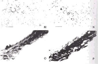
ΆΦ1 Ε‘’’ΉιΆβ≤ύΗτΚΥΡΎΝΉΥαΜ·p44/42 MAPK―υΟβ“ΏΖ¥”ΠœΗΑϊΚΆΆΜΤπΓΘ±ξ ≥Ώ Ψ40ΠΧm
ΆΦ2 Β―ιΉιΆβ≤ύΗτΚΥΡΎΝΉΥαΜ·p44/42 MAPK―υΟβ“ΏΖ¥”ΠœΗΑϊΚΆΆΜΤπΓΘ±ξ≥Ώ Ψ40ΠΧm
ΆΦ3 Ε‘’’Ήι ”«Α«χΡΎΝΉΥαΜ·p44/42 MAPK―υΟβ“ΏΖ¥”ΠœΗΑϊΚΆΆΜΤπΓΘ±ξ≥Ώ Ψ40ΠΧm
ΆΦ4 Β―ιΉι ”«Α«χΡΎΝΉΥαΜ·p44/42 MAPK―υΟβ“ΏΖ¥”ΠœΗΑϊΚΆΆΜΤπΓΘ±ξ≥Ώ Ψ40ΠΧm
ΆΦ5 Ε‘’’Ήι–”» ÷–―κΚΥΡΎΝΉΥαΜ·p44/42 MAPK―υΟβ“ΏΖ¥”ΠœΗΑϊΚΆΆΜΤπΓΘ±ξ≥Ώ Ψ40ΠΧ m
ΆΦ6 Β―ιΉι–”» ÷–―κΚΥΡΎΝΉΥαΜ·p44/42 MAPK―υΟβ“ΏΖ¥”ΠœΗΑϊΚΆΆΜΤπΓΘ±ξ≥Ώ Ψ40ΠΧm
ΆΦ7 Ε‘’’ΉιΚΘ¬μΡΎΝΉΥαΜ·p44/42 MAPK―υΟβ“ΏΖ¥”ΠœΗΑϊΚΆΆΜΤπΓΘ±ξ≥Ώ Ψ80ΠΧm
, ΑΌΡ¥“Ϋ“©
ΆΦ8 Β―ιΉιΚΘ¬μΡΎΝΉΥαΜ·p44/42 MAPK―υΟβ“ΏΖ¥”ΠœΗΑϊΚΆΆΜΤπΓΘ±ξ≥Ώ Ψ80ΠΧm
ΆΦ9 Ε‘’’Ήι “≈‘ΚΥΡΎΝΉΥαΜ·p44/42 MAPK―υΟβ“ΏΖ¥”ΠœΗΑϊΚΆΆΜΤπΓΘ±ξ≥Ώ Ψ40ΠΧm
ΆΦ10 Β―ιΉι “≈‘ΚΥΡΎΝΉΥαΜ·p44/42 MAPK―υΟβ“ΏΖ¥”ΠœΗΑϊΚΆΆΜΤπΓΘ±ξ≥Ώ Ψ40ΠΧm[ ZK)ΓΫ
ΆΦ11 Ε‘’’Ήι ”…œΚΥΡΎΝΉΥαΜ·p44/42 MAPK―υΟβ“ΏΖ¥”ΠœΗΑϊΚΆΆΜΤπΓΘ±ξ≥Ώ Ψ40ΠΧm[ ZK)ΓΫ
ΆΦ12 Β―ιΉι ”…œΚΥΡΎΝΉΥαΜ·p44/42 MAPK―υΟβ“ΏΖ¥”ΠœΗΑϊΚΆΆΜΤπΓΘ±ξ≥Ώ Ψ40ΠΧm[ ZK)ΓΫ
ΆΦ13 Ε‘’’Ήι»ιΆΖΧε…œΚΥΡΎΝΉΥαΜ·p44/42 MAPK―υΟβ“ΏΖ¥”ΠœΗΑϊΚΆΆΜΤπΓΘ±ξ≥Ώ Ψ80 ΠΧm
ΆΦ14 Β―ιΉι»ιΆΖΧε…œΚΥΡΎΝΉΥαΜ·p44/42 MAPK―υΟβ“ΏΖ¥”ΠœΗΑϊΚΆΆΜΤπΓΘ±ξ≥Ώ Ψ80 ΠΧm
, http://www.100md.com
ΆΦ15 Ε‘’’ΉιάΕΑΏΡΎΝΉΥαΜ·p44/42 MAPK―υΟβ“ΏΖ¥”ΠœΗΑϊΚΆΆΜΤπΓΘ±ξ≥Ώ Ψ40ΠΧm
ΆΦ16 Β―ιΉιάΕΑΏΡΎΝΉΥαΜ·p44/42 MAPK―υΟβ“ΏΖ¥”ΠœΗΑϊΚΆΆΜΤπΓΘ±ξ≥Ώ Ψ40ΠΧm
Fig.1 Photomicrograph showing the phosphorylated-p44/42MAPK-LI cells a nd fibers in the lateral nucleus of septum(LSV)in the control group.Bar=40ΠΧm
Fig.2 Photomicrograph showing the phosphorylated-p44/42MAPK-LI cells a nd f ibers in the lateral nucleus of septum(LSV)in the experimental group.Bar=40ΠΧm[ ZK)ΓΫ
, http://www.100md.com
Fig.3 Photomicrograph showing the phosphorylated-p44/42MAPK-LI cells a nd fibers in the preoptic area(PO)in the control group.Bar=40ΠΧm
Fig.4 Photomicrograph showing the phosphorylated-p44/42 MAPK-LI cells and fibers in the preoptic area (PO)in the experimental group.Bar=40ΠΧm
Fig.5 Photomicrograph showing the phosphorylated-p44/42 MAPK-LI cells and fibers in the central nucleus of amygdala(CeA)in the control group.Bar=40ΠΧm
, http://www.100md.com
Fig.6 Photomicrograph showing the phosphorylated-p44/42MAPK-LI cells a nd fibe rs in the central nucleus of amygdala(CeA)in the experimental group.Bar=40ΠΧm
Fig.7 Photomicrograph showing the phosphorylated-p44/42 MAPK-LI cells and fibers in the caudal hippocampus in the control group. Bar=80ΠΧm
Fig.8 Photomicrograph showing the phosphorylated-p44/42MAPK-LI cells a n d fibers in the caudal hippocampus in the experimental group.Bar=80ΠΧm
, ΑΌΡ¥“Ϋ“©
Fig.9 Photomicrograph showing the phosphorylated-p44/42MAPK-LI cells a nd fibers in the hypothalamus paraventricular nucleus(PVN)in the control group.Bar=40ΠΧm
Fig.10 Photomicrograph showing the phosphorylated-p44/42MAPK-LI cells and fiber s in the hypothalamus paraventricular nucleus(PVN)in the experimental group.Bar= 40ΠΧm
Fig.11 Photomicrograph showing the phosphorylated-p44/42MAPK-LI cells a nd fibers in the supraoptic nucleus(SON)in the control group.Bar=40ΠΧm
, http://www.100md.com
Fig.12 Photomicrograph showing the phosphorylated-p44/42MAPK-LI cells an d fibers in the supraoptic nucleus(SON) in the experimental group.Bar=40ΠΧm
Fig.13 Photomicrograph showing the phosphorylated-p44/42MAPK-LI cells an d fibers in the supramammilary nucleus(SuM)in the control group.Bar=80ΠΧm
Fig.14 Photomicrograph showing the phosphorylated-p44/42MAPK-LI cells and fibers in the supramammilary nucleus(SuM)in the experimental group.Bar=80ΠΧm
, ΑΌΡ¥“Ϋ“©
Fig.15 Photomicrograph showing the phosphorylated-p44/42MAPK-LI cells and fibers in the locus nucleus(LC)in the control group.Bar=40ΠΧm
Fig.16 Photomicrograph showing the phosphorylated-p44/42MAPK-LI cells and fibers in the locus nucleus(LC)in the experimental group.Bar=40ΠΧm
ΓΨΜυΫπœνΡΩΓΩΙζΦ“Ή‘»ΜΩΤ―ßΜυΫπΉ ÷ζœνΡΩ(39830130)ΘΜ»ΪΨϋΓΑΨ≈ΈεΓ±÷ΊΒψ ΩΈΧβΉ ÷ζœνΡΩ(96Z044)
ΓΨΉς’ΏΦρΫιΓΩΆθξΊ(1970ΓΣ)Θ§≈°(ΚΚΉε)Θ§…ΫΕΪ«ύΒΚ –»ΥΘ§ΥΕ ΩΘ§÷ς÷Έ“Ϋ Π
, http://www.100md.com
≤ΈΩΦΈΡœΉ
[1]Kortenjann MȧThomae OȧShaw PE.Inhibition of v -raf-dependent c-fos expressio n and transformation by a kinase-defective mutant of the mitogen-activated pro tein kinase Erk2[J].Mol Cell Biolȧ1994ȧ14(7):4815-4824.
[2]Huang WJȧWang BRȧYao LBȧet al.Activity of p44/42 MAP kinase in th e caudal s ubnucleus of trigeminal spinal nucleus is increased following perioral noxious s timulation in the mouse[J].Brain Resȧ2000ȧ861(1):181-185.
, ΑΌΡ¥“Ϋ“©
[3]Brunet AȧRoux DȧLenormand Pȧet al.Nuclear translocation of p42/p4 4 mitogen- activated protein kinase is required for growth factor-induced gene expression and cell cycle entry[J].EMBO Jȧ1999ȧ18(3):664-674.
[4]Alvarez EȧNorthwood ICȧGonzalez FAȧet al.Pro-Leu-Ser/Thr-Pro i s a consensus primary sequence for substrate protein phosphorylation.Characterization of the phosphorylation of c-myc and c-jun proteins by an epidermal growth factor rece pt or threonine 669 protein kinase[J].J Biol Chemȧ1991ȧ266(23):15277-15285.
, ΑΌΡ¥“Ϋ“©
[5]Miyasaka TȧMiyasaka JȧSaltiel AR.Okadaic acid stimulates the activ ity of mi crotubule associated protein kinase in PC-12 pheochromocytoma cells [J].Bioch em Biophys Res Communȧ1990ȧ168(3):1237-1243.
[6]Jovanovic JNȧBenfenati FȧSiow YLȧet al.Neurotrophins stimulate phosphory lation of synapsin I by MAP kinase and regulate synapsin I-actin interactions [J].Proc Natl Acad Sci USAȧ1996ȧ93(8):3679-3683.
[7]Atkins CMȧYon MȧGroome NPȧet al.Regulation of myelin basic protei n phosphor ylation by mitogen-activated protein kinase during increased action potential f iring in the hippocampus[J].J Neurochemȧ1999ȧ73(3):1090-1097.
, http://www.100md.com
[8]Takishima KȧGriswold PIȧIngebritsen Tȧet al.Epidermal growth fact or(EGF)rec eptor T669 peptide kinase from 3T3-L1 cells is an EGF-stimulated"MAP"kinase[J ].Proc Natl Acad Sci USAȧ1991ȧ88(6):2520-2524.
[9]Lin LLȧWartmann MȧLin AYȧet al.cPLA2 is phosphorylated and activa ted by MAP kinase [J].Cellȧ1993ȧ72(2):269-278.
[10]Kadlecova OȧMasek KȧSeifert Jȧet al The involvement of some brai n structur es in the effects of immunomodulators[J].Annals of the New York Academy of Sci enceȧ1987ȧ496(1):394-398.
, ΑΌΡ¥“Ϋ“©
[11]Nance DMȧRayson DȧCarr RIȧet al. The effects of lesions in the l ateral se ptal and hippocampal area on the humoral immune response of adult female rats[J ].Brain Behav Immunȧ1987ȧ(1):292-305.
[12]Brooks WHȧCross RJȧRoszman TLȧet al.Neuroimmunomodulation:neural anatom ical basis for impairment and facilitation[J].Ann Neurolȧ1982ȧ12(1):56-61.
[13]Rivest SȧLaflamme N.Neuronal activity and neuropeptide gene transc ription in the brains of immune-challenged rats[J].J Neuroendocrinolȧ1995ȧ7(7):501 -525.
, http://www.100md.com
[14]Saphier DȧAbramsky OȧMor Gȧet al.Multuinit electrical activity i n couci ous rats during an immune response[J].Brain Behav Immunȧ1987ȧ1(1):40-51.
[15]Lysle DTȧHoffman KEȧDykstra LA.Evidence for the involvement of th e caudal region of the periaqueductal gray in subset of morphine-induced alterations of i mmune status[J].J Pharmacol Exp Therȧ1996ȧ277(3):1533-1540.
ΓΨ ’Ηε»’ΤΎΓΩ2000-07-17 ΓΨ–όΜΊ»’ΤΎΓΩ2000-08-25, http://www.100md.com
ΒΞΈΜΘΚΒΎΥΡΨϋ“Ϋ¥σ―ß…ώΨ≠ΩΤ―ß―–ΨΩΥυ…ώΨ≠Οβ“ΏΒςΫΎ―–ΨΩ “Θ§ΈςΑ≤710032
ΙΊΦϋ¥ ΘΚΝΉΥαΜ·ΒΡp44/42 MAPKΘΜ÷§ΕύΧ«(LPS)ΘΜΡ‘ΘΜ¥σ σ
ΫβΤ ―ß±®00zk13
ΓΨ’Σ“ΣΓΩΡΩΒΡ ―–ΨΩΗΙ«ΜΉΔ…δLPSΚσ¥σ σΡ‘ΡΎΜνΜ·–Έ ΫΒΡœΗΑϊΆβ–≈Κ≈ ΒςΫΎΒΑΑΉΦΛΟΗ(p44/42 MAPK)Ζ÷≤ΦΒΡ±δΜ·ΓΘΖΫΖ®”ΟΟβ“ΏΉι÷·Μ·―ßΖ®ΓΘ ΫαΙϊ ‘ΎΗτΆβ≤ύΚΥΓΔκθœ¬«πΡ‘ΚΥΓΔ ”«Α«χΓΔ–”» ÷–―κΚΥΓΔΈ≤≤ύΚΘ¬μ œ¬«πΡ‘ “≈‘ΚΥΓΔ ”…œΚΥΓΔ»ιΆΖΧε…œΚΥΓΔ÷–―κΜ“÷ ΗΙΆβ≤ύ≤ΩΓΔ±έ≈‘Άβ≤ύΚΥΓΔάΕΑΏΒ»―τ–‘œΗΑϊ ΐΝΩ‘ωΦ” Θ§»Ψ…Ϊ‘ω«ΩΓΘΫα¬έ p44/42 MAPKΒΡΙΠΡή≤ΜΫω”κœΗΑϊΒΡ…ζ≥ΛΓΔΖ÷Μ· ΚΆ…ώΨ≠‘ΣΒΡΩ…Υή–‘±δΜ·”–ΙΊΘ§ΜΙΩ…Ρή”κΟβ“Ώ¥ΧΦΛ”–ΙΊΓΘ
ΓΨ÷–ΆΦΖ÷άύΚ≈ΓΩR322.8 ΓΨΈΡœΉ±ξ Ε¬κΓΩA ΓΨΈΡ’¬±ύΚ≈ΓΩ0529-1356(2000)-‘ω-48
, ΑΌΡ¥“Ϋ“©
THE CHANGES OF THE EXPRESSION OF PHOSPHORYLATED-p44/42 MAPK IN RAT BRAIN AFTER INTRAPERITONEAL INJECTION OF LPS
WANG XiȧWANG Bai-renȧHUANG Wen-jinȧDUAN Xiao-liȧJU Gong*
(Department of NeuroimmunomodulationΘ§Institute of NeuroscienceΘ§the F ourth Medical UniversityΘ§Xi§πan 710032Θ§China)
ΓΨAbstractΓΩObjective To study the changes of the expressio n of phosphorylated-p44/42MAPK in rat brain after intraperitoneal injection of L P S.Method Immunohistochemical staining techniques were used,an d immunostained sections were observed and analyzed under a microscope. Results The expression of phosphorylated-p44/42MAPK increased in la teral septumΘ§septohypothalamic nucleusΘ§preoptic areaΘ§central nucleus of amygd alaΘ§caudal hyppocampusΘ§hypothalamus paraventricular nucleusΘ§supraoptic nucleu sΘ§supramammilary nucleusΘ§ventrolateral part of periaqueductal grayΘ§lateral pa rabranchial nucleusΘ§locus nucleusΘ§et al.Conclusion It sugge s ted that p44/42 MAPK is related with not only cellular growthΘ§differentiation a nd plasticity of neuron but also immune stimuli probably.
, http://www.100md.com
ΓΨkey wordsΓΩp44/42MAPKΘΜ LPSΘΜ BrainΘΜ Rat ΫϋΡξά¥ΒΡ―–ΨΩ±μΟςΘ§÷– ύ…ώΨ≠œΒΆ≥Ω…“‘Ϋ” ήΟβ“Ώ–≈œΔ≤Δ≤Έ”κΕ‘Οβ“ΏΖ¥”ΠΒΡΒςΫΎΘ§ΒΪΕ‘”ΎΡ‘ΡΎ ≤Έ”κΟβ“ΏΖ¥”ΠΒΡ…ώΨ≠ΚΥΆ≈…–≤Μ«ε≥ΰΓΘ‘Ύ…ώΨ≠œΒΆ≥ΙΠΡήΚΆ–ΈΧ§œύΫαΚœΒΡ―–ΨΩ÷–Θ§Φ»Άυ”Π”ΟΫœΕύ ΒΡ «Φ¥ΩΧ‘γΤΎΜυ“ρc-fosΘ§Ά§ ±egr-1Β»“≤”–”Π”ΟΓΘ Υδ »Μ≤ΜΆ§ΒΡ”ΠΦΛΩ…“ΐΤπΡ‘ΡΎ≤ΜΆ§–Έ ΫΒΡΦ¥ΩΧ‘γΤΎΜυ“ρΒΡ±μ¥οΘ§ΒΪ «ΟΩ÷÷Φ¥ΩΧ‘γΤΎΜυ“ρ‘ΎΡ‘ΡΎΒΡ ±μ¥οΨυ”–“ΜΕ®ΒΡΨ÷œό–‘ΓΘ“ρ¥ΥΘ§–η“ΣΫαΚœΕύ÷÷±ξ÷ΨΈο≤≈ΡήΗϋ»ΪΟφΒΊΖ¥”≥≥ω≤Έ”κΟβ“ΏΖ¥”ΠΒΡΡ‘ ΡΎ…ώΨ≠ΚΥΆ≈ΒΡΜνΕ·«ιΩωΓΘ±Ψ―–ΨΩ¥”–≈Κ≈ΉΣΒΦΖ÷Ή”ΒΡΜΖΫΎ―–ΨΩΝΥΟβ“ΏΖ¥”Π ±…ώΨ≠ΚΥΆ≈ΒΡΜνΕ·«ι ΩωΓΘ
œΗΑϊΆβ–≈Κ≈ΒςΫΎΒΑΑΉΦΛΟΗ(p44/42 MAPKΘ§Φ¥ERK1/ERK2) τ”ΎΥΩΝ―‘≠ΜνΜ·ΒΑΑΉΦΛΟΗ(MAPK)Φ“Ήε Θ§ «“Μάύ÷Ί“ΣΒΡ–≈Κ≈ΉΣΒΦΖ÷Ή”Θ§Υϋ±ΜΦΛΜν≥…ΈΣΝΉΥαΜ·ΒΡp44/42 MAPKΖΔΜ”Ής”ΟΘ§≤Έ”κΒςΫΎœΗ ΑϊΒΡ”–ΥΩΖ÷Ν―ΚΆΖ÷Μ·[1]Θ§≤Δ‘Ύ…ώΨ≠‘ΣΚΆΫΚ÷ œΗΑϊΒΡ–≈Κ≈¥ΪΒί÷–ΤπΉ≈÷Ί“ΣΒΡΉς”ΟΓΘ Έ“Ο«[2]–¬ΫϋΒΡ―–ΨΩΖΔœ÷Θ§»ΐ≤φ…ώΨ≠ΦΙ χΚΥΈ≤≤ύ―«ΚΥΒΡp44/42 MAPKΜν–‘‘ΎΟφ≤Ω…ΥΚΠ –‘¥ΧΦΛ ±±μ¥ο…œΒςΘ§‘Ύ±Ψ―–ΨΩ÷–Έ“Ο«”ΟΟβ“ΏΉι÷·Μ·―ßΖΫΖ®Θ§―–ΨΩΝΥΗΙ«ΜΡΎΗχ”ηΟβ“Ώ¥ΧΦΛΦΝ÷§ ΕύΧ«(lipopolysacchrideΘ§LPS)ΚσΡ‘ΡΎΝΉΥαΜ·ΒΡp44/42 MAPK±μ¥οΒΡ±δΜ·ΓΘ
, http://www.100md.com
≤ΡΝœΚΆΖΫΖ®
1. Ε·ΈοΉΦ±ΗΚΆ«–Τ§ΒΡ÷Τ±Η
–έ–‘SD¥σ σ8÷ΜΘ§Χε÷Ί180ΓΪ250gΘ§ΥφΜζΖ÷ΈΣ2ΉιΘΚ Β―ιΉι4÷ΜΘ§ΗΙ«ΜΡΎΉΔ…δLPS(500ΠΧg/kgΘΜS igmaΙΪΥΨΘΜ”Ο…ζάμ―ΈΥ°œΓ ΆΈΣ100mg/L)ΘΜΕ‘’’Ήι4÷ΜΘ§ΟΩΙΪΫοΧε÷ΊΗΙ«ΜΡΎΉΔ…δΆ§ΧεΜΐΒΡ…ζάμ ―ΈΥ°ΓΘ3hΚσΘ§1%ΈλΑΆ±»ΆΉΡΤ(40mg/kg)ΗΙ«ΜΉΔ…δΘ§¬ιΉμΚσΩΣ–ΊΘ§Ψ≠–ΡΙύΝςΘ§œ»”Ο100ml…ζάμ―Έ Υ°ΩλΥΌΙύΉΔ≥εœ¥―Σ“ΚΘ§»ΜΚσ”Ο‘ΛάδΒΡ4%ΕύΨέΦΉ»©ΙΧΕ®“Κ400mlΙύΝςΓΘ¥ΐΙΧΕ®“ΚΒΈΉΔΆξ±œΘ§»Γ ≥ω»ΪΡ‘Θ§”Ο4%ΕύΨέΦΉ»©ΚσΙΧΕ®2hΘ§‘ΌΫΪΡ‘Ζ≈»κ20%’αΧ«»ή“ΚΘ§4ΓφΙΐ“ΙΓΘΉι÷·ΩιΆξ»Ϊ≥ΝΒΉΚσ”Ο ±υΕ≥«–Τ§ΜζΝ§–χΙΎΉ¥«–Τ§Θ§ΟΩ5’≈»Γ1’≈Θ§Τ§Κώ50ΠΧmΓΘ
2.Οβ“ΏΉι÷·Μ·―ß»Ψ…Ϊ
œ»ΫΪ«–Τ§Ψ≠0.01mol/L PBSΤ·œ¥5minΓΝ3¥ΈΚσΘ§Ϋΰ»κ80%ΦΉ¥ΦΚΆ0.3%Ιΐ―θΜ·«βΖβ±’30minΘ§0 .01mol/L PBSΤ·œ¥10minΓΝ3¥ΈΘ§‘Ό”Ο1%≈Θ―Σ«εΑΉΒΑΑΉΓΔ3%―ρ―Σ«εΚΆ0.3% Triton X-100Ζβ ±’1hΘ§Ά§«ΑΤ·œ¥ΙΐΚσΘ§”ΟABCΖΫΖ®Ϋχ––Οβ“ΏΉι÷·Μ·―ß»Ψ…ΪΘΚΆΟΩΙ¥σ σΝΉΥαΜ·p44/42 MAPKΩΙ Χε(1ΘΚ500Θ§NEB ΙΪΥΨ)Ζθ”ΐ«–Τ§Θ§ “Έ¬œ¬36hΘ§0.01mol/L PBS Τ·œ¥10minΓΝ3¥ΈΘΜ…ζΈοΥΊΜ· ΒΡ―ρΩΙΆΟIgG(1:500Θ§SigmaΙΪΥΨ)Ζθ”ΐ«–Τ§Θ§ “Έ¬œ¬4hΘ§0.01mol/L PBSΤ·œ¥10minΓΝ3¥Έ ΘΜ…ζΈοΥΊ-¬―ΑΉΥΊ-ά±ΗυΙΐ―θΜ·ΈοΟΗΗ¥ΚœΈο(ABCΘ§1ΘΚ500Θ§ VectorΙΪΥΨ)Ζθ”ΐ«–Τ§Θ§ “Έ¬2h Θ§ 0.01mol/L PBSΤ·œ¥10minΓΝ3¥ΈΘΜΝρΥαΡχΑΖΦ”«ΩDABάΕ…ΪΖ¥”Π≥ …ΪΓΘΒ±―τ–‘≤ζΈο≥ …νάΕ…ΪΕχ ±≥ΒΉΦΗΚθΈό…Ϊ ±÷’÷ΙΖ¥”ΠΓΘΨ≠Ά―Υ°ΓΔΆΗΟςΚΆΖβΤ§ΚσΘ§ΨΒΦλΓΘ
, ΑΌΡ¥“Ϋ“©
≤ΩΖ÷«–Τ§”ΟΉωΟβ“ΏΉι÷·Μ·―ßΖΫΖ®Ε‘’’ Β―ιΘ§“ΜΩΙ”Ο≈Θ―Σ«εΑΉΒΑΑΉœΓ Ά“ΚΧφ¥ζΘ§ΕΰΩΙΓΔABCΗ¥ ΚœΈοΆ§ΤδΥϊΟβ“ΏΉι÷·Μ·―ßΖ¥”Π«–Τ§Θ§ΫαΙϊΈ¥Φϊ―τ–‘ΫαΙΙΓΘ
3.ΝΉΥαΜ·p44/42 MAPK―τ–‘œΗΑϊΦΤ ΐΖΫΖ®
ΗυΨίΚΥΆ≈ΒΡ¥σ–ΓΘ§»ΓΟΩ÷ΜΕ·ΈοΒΡΟΩΗωΚΥΆ≈œύΕ‘”ΠΒΡΒδ–ΆΤΫΟφ1ΓΪ2ΗωΘ§‘Ύ10ΓΝ20ΙβΨΒœ¬Ιέ≤λ≤Δ ΦΤ ΐ―τ–‘œΗΑϊ ΐ ΓΘΗΟάΐΕ·ΈοΗΟΚΥΆ≈ΒΡΤΫΨυ ΐΘ§Φ¥ΈΣΗΟΕ·ΈοΗΟΚΥΆ≈ΒΡ―τ–‘œΗΑϊ ΐΘΜΟΩΉι4ΗωΕ·ΈοΦδ»ΓΤΫΨυ÷ΒΘ§ Φ¥ΈΣΗΟΉιΕ·ΈοΗΟΚΥΆ≈ΒΡ―τ–‘œΗΑϊ ΐΓΘ≤…”ΟtΦλ―ιΫχ––Ά≥ΦΤΖ÷ΈωΓΘ
Ϋα Ιϊ
Ε‘ Β―ιΉιΚΆΕ‘’’Ήι¥”―”ΥηΈ≤ΕΥΒΫΕΥΡ‘ΒΡ»ΪΡ‘«–Τ§Ϋχ––ΝΥΕ‘±»Ιέ≤λΘ§―τ–‘≤ζΈοΦϊ”Ύ…ώΨ≠‘ΣΒΡΑϊ Ϋ§ΓΔΑϊΚΥΚΆΆΜΤπΘ§¥φ‘Ύ”Ύ…ώΨ≠‘ΣΒΡΑϊΧεΓΔ ςΆΜΚΆ÷αΆΜ÷–ΒΡΝΩΟςœ‘Εύ”ΎœΗΑϊΚΥ÷–ΒΡΝΩΓΘ‘ΎΕ‘’’ ΉιΕ·ΈοΘ§ΜνΜ·ΒΡp44/42MAPK≥ωœ÷‘Ύ–μΕύΚΥΆ≈Θ§ΧΊ±π «‘ΎΒΚΤΛ÷ ΓΔΗ–Ψθ‘ΥΕ·ΤΛ÷ ΚΆ ”ΓΔΧΐΤΛ÷ ΓΔΚΘ¬μΡΎ–α«χΚΆ±≥≤ύΓΔΗΙ≤ύœ¬Ά–Θ§ΡΎ≤ύ–”» ΚΥΓΔΤΛ÷ –”» ΚΥΚΆ÷–ΫιΚΥΘ§ ”ΫΜ≤φ…œΚΥΓΔœ¬«πΡ‘ “≈‘ΚΥ¥σœΗΑϊ≤ΩΓΔΙ≠Ή¥ΚΥΓΔ«ΑΗΙ≤ύ ”«ΑΚΥΓΔΆβ≤ύ«ΑΚΥΓΔ»ιΆΖΧε…œΚΥΚΆœ¬«πΡ‘Κσ«χΓΔΕΞΗ««Α«χ ΓΔ÷–―κΜ“÷ ΗΙΆβ≤ύ≤ΩΓΔάΕΑΏΓΔ±έ≈‘Άβ≤ύΚΥΓΔ–ΓΡ‘Τ÷Ωœ“ΑœΗΑϊΓΔA5«χΓΔ≈‘ΨόœΗΑϊΆβ≤ύ ΚΥΓΔΈ«≤ύ(C1«χ)ΚΆΈ≤≤ύ(A1«χ)―”ΥηΗΙΆβ≤ύΆχΉ¥ΫαΙΙΒ»ΓΘ
, http://www.100md.com
Β―ιΉιΚΆΕ‘’’ΉιΒΡ≤ΜΆ§Βψ «Θ§‘ΎΡ≥–©Ρ‘«χΘ§ΝΉΥαΜ·p44/42 MAPK―υΟβ“ΏΖ¥”Π…ώΨ≠‘ΣΒΡ ΐΝΩ ΦΑ ―τ–‘…ώΨ≠œΥΈ§ΒΡΟήΕ»ΚΆ≥ΛΕ»ΖΔ…ζΗΡ±δΓΘ”κΕ‘’’Ήι±»ΫœΘ§ Β―ιΉι‘ΎΆβ≤ύΗτ«χ(ΆΦ1Θ§2)ΓΔΗτœ¬«π Ρ‘ΚΥΓΔ ”«Α«χ(ΆΦ3Θ§4)ΓΔ–”» ÷–―κΚΥ(ΆΦ5Θ§6)ΓΔΈ≤≤ύΚΘ¬μ(ΆΦ7Θ§8)ΓΔœ¬«πΡ‘ “≈‘ΚΥ(ΆΦ9Θ§10 )ΓΔ ”…œΚΥ(ΆΦ11Θ§12)ΓΔ»ιΆΖΧε…œΚΥ(ΆΦ13Θ§14)ΓΔ÷–―κΜ“÷ ΗΙΆβ≤ύ≤ΩΓΔΆβ≤ύ±έ≈‘ΚΥΓΔάΕΑΏ(ΆΦ 15Θ§16)¥Π―τ–‘œΗΑϊ ΐΝΩ‘ωΦ”ΓΔ»Ψ…Ϊ‘ω«ΩΓΘ ΐΝΩ±»ΫœΦϊΗΫ±μΓΘ
ΗΫ±μ Β―ιΉι(n=4)ΚΆΕ‘’’Ήι(n =4)Ρ‘ΡΎ≤Ω
Ζ÷ΚΥΆ≈÷–ΝΉΥαΜ·p44/42 MAPK―τ–‘œΗΑϊΦΤ ΐ
n=4)and experimental group(n=4) ΚΥΆ≈Οϊ≥Τ
Ε‘’’ Ήι
Β―ιΉι
, ΑΌΡ¥“Ϋ“©
nuclei
control group
experimental group
Άβ≤ύΗτ«χ
25Γά4
48Γά7a
(lateral septal region)
Ητœ¬«πΡ‘ΚΥ
31Γά9
76Γά13a
(septohypothalamic nucleus)
, ΑΌΡ¥“Ϋ“©
”«Α«χ
18Γά3
45Γά7a
(preoptic region)
–”» ÷–―κΚΥ
19Γά5
83Γά11a
(central amygdaloid nuclei)
Έ≤≤ύΚΘ¬μ
6Γά2
21Γά4a
, ΑΌΡ¥“Ϋ“©
(caudal hippocampus)
œ¬«πΡ‘ “≈‘ΚΥ
69Γά0
120Γά19a
(hypothalamic paraventricular nucleus)
”…œΚΥΚσ≤Ω
21Γά13
128Γά23a
(retrochiasmatic part of supraoptic nucleus)
»ιΆΖΧε…œΚΥ
, http://www.100md.com
38Γά8
126Γά31a
(supramammillary nucleus)
÷–―κΜ“÷ ΗΙΆβ≤ύ≤Ω
13Γά7
72Γά19a
(lateral ventral part of central gray)
Άβ≤ύ±έ≈‘ΚΥ
28Γά8
59Γά12a
, http://www.100md.com (lateral parabrachial nucleus)
άΕΑΏ
54Γά9
93Γά13a
(locus ceruleus)
a.”κΕ‘’’Ήιœύ±»P<0.05 a.P<0.05 vs.Contr ol group.
Χ÷ ¬έ
ΥΩΝ―‘≠ΜνΜ·ΒΑΑΉΦΛΟΗ(MAPK) τœΗΑϊΡΎΒΑΑΉΥΩΑ±Υα/Υ’Α±ΥαΦΛΟΗΦ“Ήε≥…‘±Θ§ «“Μάύ÷Ί“ΣΒΡΫ” ή ΒΞ¥ΈΩγΡΛ ήΧεΉΣΜΜ”κ¥ΪΒίΒΡ–≈Κ≈≤ΔΫΪΤδ¥χ»κœΗΑϊΚΥΡΎΒΡ–≈Κ≈ΉΣΒΦΖ÷Ή”Θ§≤Έ”κΕύ÷÷œΗΑϊΙΠΡήΒΡ ΒςΩΊΓΘΤυΫώΈΣ÷ΙΘ§‘ΎMAPKΦ“Ήε÷–“―”–3Ηω―«Φ“Ήε±ΜΟη ωΘ§Ζ÷±πΈΣp44/42 MAPK(ERK1/ERK2Θ§œΗ ΑϊΆβ–≈Κ≈ΒςΫΎΒΑΑΉΦΛΟΗ)ΓΔJNK(c-junNΡ©ΕΥΒΑΑΉΦΛΟΗΘ§“≤±Μ≥ΤΈΣSAPKΘ§Φ¥”Π Φ±ΦΛΜνΒΑΑΉΦΛΟΗ)ΚΆp38 MAPK(“≤±Μ≥ΤΉςHOGΘ§Φ¥ΗΏ…χΆΗ–‘Η ”ΆΖ¥”ΠΦΛΟΗΓΘ)…ζ≥Λ“ρΉ”ΚΆ”–ΥΩΖ÷ Ν―–≈Κ≈Ά®ΙΐRasΓΔRafΓΔMEK1/MEK2Β»“ΜœΒΝ–ΦΕΝ§Ζ¥”Π Ιp44/42 MAPKΝΉΥαΜ·Θ§≥…ΈΣΜνΜ·ΒΡp44/ 42 MAPKΕχΖΔΜ”Ής”ΟΓΘ
, ΑΌΡ¥“Ϋ“©
p42/44 MAPK±ΜΦΛΜνΚσΘ§Φ»Ω…“‘¥”ΑϊΫ§Ϋχ»κΑϊΚΥ≤ΔΉς”Ο”ΎΚΥΒΑΑΉ»γElk1ΓΔc-mycΚΆTAL1 [ 3Θ§4]Β»ΉΣ¬Φ“ρΉ”Θ§”Αœλœ¬”ΈΜυ“ρΒΡ±μ¥οΘ§”÷Ω…“‘‘ΎΑϊΫ§ΡΎΉς”Ο”ΎΒΉΈοΥΩΝ―‘≠ΜνΜ·ΒΑΑΉ- 2 (MAP2)[5]ΓΔΆΜ¥ΞΥΊ[6]ΓΔΥη« Φν–‘ΒΑΑΉ(MBP)[7]ΜρœΗΑϊ±μΟφΖ÷Ή” »γ±μΤΛ…ζ≥Λ“ρΉ” ήΧε[8]ΓΔΝΉ÷§ΟΗA2[9]Θ§≤Δ ΙΒΉΈοΝΉΥαΜ·Θ§”ΑœλœΗΑϊΙ«Φή ΒΡ÷ΊΥήΚΆΆΜ¥Ξ–Έ≥…ΓΘ±Ψ Β―ι÷–Θ§Έ“Ο«ΖΔœ÷ΜνΜ·ΒΡp44/42 MAPK¥φ‘Ύ”Ύ…ώΨ≠‘ΣΒΡΑϊΧεΓΔ ςΆΜΚΆ ÷α ΆΜ÷–ΒΡΝΩΟςœ‘Εύ”ΎœΗΑϊΚΥ÷–ΒΡΝΩΓΘΥδ»ΜΝΉΥαΜ·ΒΡp44/42 MAPKΩ…“‘¥”ΑϊΫ§ΉΣ“ΤΒΫΚΥΡΎΘ§ ΙΚΥ ΡΎ ΒΡΉΣ¬Φ“ρΉ”ΝΉΥαΜ·Θ§ΒΪ «÷αΆΜΚΆ ςΆΜ÷–ΒΡΝΉΥαΜ·ΒΡp44/42 MAPKΨύάκœΗΑϊΚΥΫœ‘ΕΘ§Υυ“‘ΝΉΥα Μ· ΒΡp44/42 MAPKΗϋ”–Ω…ΡήΉς”Ο”ΎΑϊΫ§÷–ΒΡΒΉΈοΘ§±»»γMAP2ΓΔTALΓΔMBPΚΆΆΜ¥ΞΥΊΘ§≤Έ”κΟβ“Ώ”Π ΦΛ“ΐΤπ…ώΨ≠‘ΣΙΠΡή¥ζ–ΜΚσΖΔ…ζΒΡœΗΑϊΙ«ΦήΚΆΆΜ¥ΞΒΡ÷ΊΥήΓΘ
±Ψ―–ΨΩΫαΙϊœ‘ Ψ‘ΎΕ‘’’Ήι¥σ σΡ‘÷–Θ§ΜνΜ·ΒΡp44/42 MAPK”–ΙψΖΚΒΡ±μ¥οΘ§’βΧα ΨΝΥp44/42 MA PK‘ΎΡ‘ΒΡ÷ΎΕύΙΠΡήΒΡ–≈Κ≈¥ΪΒΦΙΐ≥Χ÷–ΒΡ÷Ί“Σ–‘ΓΘ‘ΎΈ“Ο«“‘ΆυΒΡ Β―ι÷–Θ§ΖΔœ÷Ηχ”ηΕ·ΈοΧέΆ¥¥Χ ΦΛΚσΘ§Ρ‘ΡΎ»ΐ≤φ…ώΨ≠ΦΙ χΚΥΈ≤≤ύ―«ΚΥΝΉΥαΜ·ΒΡp44/42 MAPK±μ¥ο‘ωΗΏ[2]Θ§Χα Ψp44/ 42 MAPKΒΡΙΠΡή‘Ε≤Μ÷Ι“‘ΆυΈΡœΉ÷–ΥυΧαΒΫΒΡΡ«–©ΓΘ“ρ¥ΥΘ§±Ψ―–ΨΩ÷–Έ“Ο«”ΟΟβ“ΏΉι÷·Μ·―ßΖΫΖ® Θ§Ιέ ≤λΝΥΗΙ«ΜΡΎΗχ”ηΟβ“Ώ¥ΧΦΛΦΝLPSΚσΡ‘ΡΎΝΉΥαΜ·ΒΡp44/42 MAPK±μ¥οΒΡ±δΜ·Θ§ΫαΙϊΖΔœ÷Ρ‘ΡΎΝΉΥα Μ·ΒΡp44/42 MAPK±μ¥ο‘ωΦ”ΓΘ
, ΑΌΡ¥“Ϋ“©
Φ»Άυ―–ΨΩΖΔœ÷Θ§ΥπΜΌάΕΑΏΚΆ±έ≈‘ΚΥΒ»Ρ‘Η…ΫαΙΙΨυ≤ΜΆ§≥ΧΕ»ΒΊ”ΑœλΟβ“ΏΙΠΡή[10]ΘΜœ¬ «πΡ‘“Μ÷±±Μ»œΈΣ «…ώΨ≠-ΡΎΖ÷ΟΎ-Οβ“ΏΒςΫΎΒΡ÷– ύΘ§ΗτΆβ≤ύΚΥ”κœ¬«πΡ‘ΙΊœΒΟή«–Θ§ΗτΆβ≤ύΚΥ Υπ ΜΌΩ…“ΐΤπTœΗΑϊ”Π¥πΒΡΟςœ‘ΗΡ±δ[11]ΘΜΥπΜΌ±≥≤ύΚΘ¬μΚΆ–”» Η¥ΚœΧεΒΦ÷¬“ΜΙΐ–‘ΒΡΤΔ ΓΔ–ΊœΌœΗΑϊ ΐΝΩ‘ωΦ”ΚΆTœΗΑϊ‘ω÷≥Ζ¥”Π‘ω«Ω[12]ΘΜRivest[13]ΖΔœ÷ΗΙ«ΜΡΎ ΉΔ…δLPSΘ§3hΚσ“‘œ¬ΚΥΆ≈ΒΡc-fosmRNA¥οΒΫ±μ¥οΗΏΖεΘΚ “≈‘ΚΥ¥σœΗΑϊ≤ΩΚΆ–Γ œΗΑϊ≤ΩΓΔ ”…œΚΥΓΔΡΎ≤ύ ”«Α«χΓΔάΕΑΏΓΔ–”» ÷–―κΚΥΚΆΆβ≤ύ±έ≈‘ΚΥΒ»ΓΘSaphierΒ»[14] ÷ΛΟςΘ§SRBCsΟβ“ΏΕ·ΈοΚσΘ§‘Ύ≥θ¥ΈΩΙΧε…ζ≥…ΗΏΖεΤΎ(Οβ“ΏΚσΒΎ5d)PO/AHΖ≈Βγ‘ωΦ”Θ§”ΟΜΖΑϊΨζ ΥΊA“÷÷ΤΆβ÷ήΟβ“ΏΖ¥”ΠΚσΘ§÷– ύΒΡΖ≈Βγ±δΜ·œϊ ßΘ§Χα ΨPO/AHΒΡΖ≈Βγ±δΜ·”κΆβ÷ήΟβ“ΏΖ¥”Π”–ΙΊ ΓΘ LysleΒ»[15]±®ΒάΘ§ΫΪ¬πΖ»ΉΔ…δΒΫ÷–―κΜ“÷ ΗΙΆβ≤ύ≤ΩΘ§Ω…“ΐΤπTΓΔBΝήΑΆœΗΑϊ‘ω÷≥ΙΠ ΡήΫΒΒΆΓΔΉ‘»Μ…±…ΥœΗΑϊΕΨ–‘ΫΒΒΆΓΔIL-2ΚΆΠΟ-Η…»≈ΥΊΚœ≥…Φθ…ΌΓΘ“‘…œ±®ΒάΨυΧα Ψ’β–©ΚΥΆ≈ ”κΟβ“ΏΒςΫΎ”–ΙΊΘ§Εχ’β–©ΚΥΆ≈‘Ύ±Ψ Β―ι÷–Οβ“Ώ¥ΧΦΛΚσΝΉΥαΜ·p44/42 MAPK±μ¥οΨυ‘ωΦ”ΓΔ‘ω«Ω Θ§ Χα Ψp44/42 MAPK≤Έ”κΝΥ’β–©ΚΥΆ≈Ε‘Οβ“ΏΖ¥”ΠΒΡΒςΫΎΓΘΈΡœΉ±®ΒάLPSΗΙ«ΜΡΎΉΔ…δΚσΘ§¥ΧΦΛΗΙ «ΜΡΎΨό …œΗΑϊ≤ζ…ζIL-1Π¬ΓΔTNF-ΠΝΚΆIL-6Β»¥Ό―Ή–‘œΗΑϊ“ρΉ”ΓΘΈ“Ο«ΆΤ≤β’β–©œΗΑϊ“ρΉ”Φ» Ω…Ρή Ά®Ιΐ―Σ‘¥–‘ΆΨΨΕΫχ»κΡ‘≤ΔΉς”Ο”ΎΡ‘Θ§”÷Ω…ΡήΆ®Ιΐ…ώΨ≠Μζ÷ΤΫΪΟβ“Ώ–≈œΔ¥ΪΒίΒΫΡ‘Θ§ ΙΡ‘ΡΎ≤ζ…ζ œΗΑϊ“ρΉ”ΜρΤδΥϊΫι÷ Θ§Ά®ΙΐMAPKΆΨΨΕ”Αœλ≤ΜΆ§ΚΥΆ≈ΡΎ…ώΨ≠‘ΣΒΡΙΠΡήΜνΕ·ΓΘ
, ΑΌΡ¥“Ϋ“©
ΆΦ1 Ε‘’’ΉιΆβ≤ύΗτΚΥΡΎΝΉΥαΜ·p44/42 MAPK―υΟβ“ΏΖ¥”ΠœΗΑϊΚΆΆΜΤπΓΘ±ξ ≥Ώ Ψ40ΠΧm
ΆΦ2 Β―ιΉιΆβ≤ύΗτΚΥΡΎΝΉΥαΜ·p44/42 MAPK―υΟβ“ΏΖ¥”ΠœΗΑϊΚΆΆΜΤπΓΘ±ξ≥Ώ Ψ40ΠΧm
ΆΦ3 Ε‘’’Ήι ”«Α«χΡΎΝΉΥαΜ·p44/42 MAPK―υΟβ“ΏΖ¥”ΠœΗΑϊΚΆΆΜΤπΓΘ±ξ≥Ώ Ψ40ΠΧm
ΆΦ4 Β―ιΉι ”«Α«χΡΎΝΉΥαΜ·p44/42 MAPK―υΟβ“ΏΖ¥”ΠœΗΑϊΚΆΆΜΤπΓΘ±ξ≥Ώ Ψ40ΠΧm
ΆΦ5 Ε‘’’Ήι–”» ÷–―κΚΥΡΎΝΉΥαΜ·p44/42 MAPK―υΟβ“ΏΖ¥”ΠœΗΑϊΚΆΆΜΤπΓΘ±ξ≥Ώ Ψ40ΠΧ m
ΆΦ6 Β―ιΉι–”» ÷–―κΚΥΡΎΝΉΥαΜ·p44/42 MAPK―υΟβ“ΏΖ¥”ΠœΗΑϊΚΆΆΜΤπΓΘ±ξ≥Ώ Ψ40ΠΧm
ΆΦ7 Ε‘’’ΉιΚΘ¬μΡΎΝΉΥαΜ·p44/42 MAPK―υΟβ“ΏΖ¥”ΠœΗΑϊΚΆΆΜΤπΓΘ±ξ≥Ώ Ψ80ΠΧm
, ΑΌΡ¥“Ϋ“©
ΆΦ8 Β―ιΉιΚΘ¬μΡΎΝΉΥαΜ·p44/42 MAPK―υΟβ“ΏΖ¥”ΠœΗΑϊΚΆΆΜΤπΓΘ±ξ≥Ώ Ψ80ΠΧm
ΆΦ9 Ε‘’’Ήι “≈‘ΚΥΡΎΝΉΥαΜ·p44/42 MAPK―υΟβ“ΏΖ¥”ΠœΗΑϊΚΆΆΜΤπΓΘ±ξ≥Ώ Ψ40ΠΧm
ΆΦ10 Β―ιΉι “≈‘ΚΥΡΎΝΉΥαΜ·p44/42 MAPK―υΟβ“ΏΖ¥”ΠœΗΑϊΚΆΆΜΤπΓΘ±ξ≥Ώ Ψ40ΠΧm[ ZK)ΓΫ
ΆΦ11 Ε‘’’Ήι ”…œΚΥΡΎΝΉΥαΜ·p44/42 MAPK―υΟβ“ΏΖ¥”ΠœΗΑϊΚΆΆΜΤπΓΘ±ξ≥Ώ Ψ40ΠΧm[ ZK)ΓΫ
ΆΦ12 Β―ιΉι ”…œΚΥΡΎΝΉΥαΜ·p44/42 MAPK―υΟβ“ΏΖ¥”ΠœΗΑϊΚΆΆΜΤπΓΘ±ξ≥Ώ Ψ40ΠΧm[ ZK)ΓΫ
ΆΦ13 Ε‘’’Ήι»ιΆΖΧε…œΚΥΡΎΝΉΥαΜ·p44/42 MAPK―υΟβ“ΏΖ¥”ΠœΗΑϊΚΆΆΜΤπΓΘ±ξ≥Ώ Ψ80 ΠΧm
ΆΦ14 Β―ιΉι»ιΆΖΧε…œΚΥΡΎΝΉΥαΜ·p44/42 MAPK―υΟβ“ΏΖ¥”ΠœΗΑϊΚΆΆΜΤπΓΘ±ξ≥Ώ Ψ80 ΠΧm
, http://www.100md.com
ΆΦ15 Ε‘’’ΉιάΕΑΏΡΎΝΉΥαΜ·p44/42 MAPK―υΟβ“ΏΖ¥”ΠœΗΑϊΚΆΆΜΤπΓΘ±ξ≥Ώ Ψ40ΠΧm
ΆΦ16 Β―ιΉιάΕΑΏΡΎΝΉΥαΜ·p44/42 MAPK―υΟβ“ΏΖ¥”ΠœΗΑϊΚΆΆΜΤπΓΘ±ξ≥Ώ Ψ40ΠΧm
Fig.1 Photomicrograph showing the phosphorylated-p44/42MAPK-LI cells a nd fibers in the lateral nucleus of septum(LSV)in the control group.Bar=40ΠΧm
Fig.2 Photomicrograph showing the phosphorylated-p44/42MAPK-LI cells a nd f ibers in the lateral nucleus of septum(LSV)in the experimental group.Bar=40ΠΧm[ ZK)ΓΫ
, http://www.100md.com
Fig.3 Photomicrograph showing the phosphorylated-p44/42MAPK-LI cells a nd fibers in the preoptic area(PO)in the control group.Bar=40ΠΧm
Fig.4 Photomicrograph showing the phosphorylated-p44/42 MAPK-LI cells and fibers in the preoptic area (PO)in the experimental group.Bar=40ΠΧm
Fig.5 Photomicrograph showing the phosphorylated-p44/42 MAPK-LI cells and fibers in the central nucleus of amygdala(CeA)in the control group.Bar=40ΠΧm
, http://www.100md.com
Fig.6 Photomicrograph showing the phosphorylated-p44/42MAPK-LI cells a nd fibe rs in the central nucleus of amygdala(CeA)in the experimental group.Bar=40ΠΧm
Fig.7 Photomicrograph showing the phosphorylated-p44/42 MAPK-LI cells and fibers in the caudal hippocampus in the control group. Bar=80ΠΧm
Fig.8 Photomicrograph showing the phosphorylated-p44/42MAPK-LI cells a n d fibers in the caudal hippocampus in the experimental group.Bar=80ΠΧm
, ΑΌΡ¥“Ϋ“©
Fig.9 Photomicrograph showing the phosphorylated-p44/42MAPK-LI cells a nd fibers in the hypothalamus paraventricular nucleus(PVN)in the control group.Bar=40ΠΧm
Fig.10 Photomicrograph showing the phosphorylated-p44/42MAPK-LI cells and fiber s in the hypothalamus paraventricular nucleus(PVN)in the experimental group.Bar= 40ΠΧm
Fig.11 Photomicrograph showing the phosphorylated-p44/42MAPK-LI cells a nd fibers in the supraoptic nucleus(SON)in the control group.Bar=40ΠΧm
, http://www.100md.com
Fig.12 Photomicrograph showing the phosphorylated-p44/42MAPK-LI cells an d fibers in the supraoptic nucleus(SON) in the experimental group.Bar=40ΠΧm
Fig.13 Photomicrograph showing the phosphorylated-p44/42MAPK-LI cells an d fibers in the supramammilary nucleus(SuM)in the control group.Bar=80ΠΧm
Fig.14 Photomicrograph showing the phosphorylated-p44/42MAPK-LI cells and fibers in the supramammilary nucleus(SuM)in the experimental group.Bar=80ΠΧm
, ΑΌΡ¥“Ϋ“©
Fig.15 Photomicrograph showing the phosphorylated-p44/42MAPK-LI cells and fibers in the locus nucleus(LC)in the control group.Bar=40ΠΧm
Fig.16 Photomicrograph showing the phosphorylated-p44/42MAPK-LI cells and fibers in the locus nucleus(LC)in the experimental group.Bar=40ΠΧm
ΓΨΜυΫπœνΡΩΓΩΙζΦ“Ή‘»ΜΩΤ―ßΜυΫπΉ ÷ζœνΡΩ(39830130)ΘΜ»ΪΨϋΓΑΨ≈ΈεΓ±÷ΊΒψ ΩΈΧβΉ ÷ζœνΡΩ(96Z044)
ΓΨΉς’ΏΦρΫιΓΩΆθξΊ(1970ΓΣ)Θ§≈°(ΚΚΉε)Θ§…ΫΕΪ«ύΒΚ –»ΥΘ§ΥΕ ΩΘ§÷ς÷Έ“Ϋ Π
, http://www.100md.com
≤ΈΩΦΈΡœΉ
[1]Kortenjann MȧThomae OȧShaw PE.Inhibition of v -raf-dependent c-fos expressio n and transformation by a kinase-defective mutant of the mitogen-activated pro tein kinase Erk2[J].Mol Cell Biolȧ1994ȧ14(7):4815-4824.
[2]Huang WJȧWang BRȧYao LBȧet al.Activity of p44/42 MAP kinase in th e caudal s ubnucleus of trigeminal spinal nucleus is increased following perioral noxious s timulation in the mouse[J].Brain Resȧ2000ȧ861(1):181-185.
, ΑΌΡ¥“Ϋ“©
[3]Brunet AȧRoux DȧLenormand Pȧet al.Nuclear translocation of p42/p4 4 mitogen- activated protein kinase is required for growth factor-induced gene expression and cell cycle entry[J].EMBO Jȧ1999ȧ18(3):664-674.
[4]Alvarez EȧNorthwood ICȧGonzalez FAȧet al.Pro-Leu-Ser/Thr-Pro i s a consensus primary sequence for substrate protein phosphorylation.Characterization of the phosphorylation of c-myc and c-jun proteins by an epidermal growth factor rece pt or threonine 669 protein kinase[J].J Biol Chemȧ1991ȧ266(23):15277-15285.
, ΑΌΡ¥“Ϋ“©
[5]Miyasaka TȧMiyasaka JȧSaltiel AR.Okadaic acid stimulates the activ ity of mi crotubule associated protein kinase in PC-12 pheochromocytoma cells [J].Bioch em Biophys Res Communȧ1990ȧ168(3):1237-1243.
[6]Jovanovic JNȧBenfenati FȧSiow YLȧet al.Neurotrophins stimulate phosphory lation of synapsin I by MAP kinase and regulate synapsin I-actin interactions [J].Proc Natl Acad Sci USAȧ1996ȧ93(8):3679-3683.
[7]Atkins CMȧYon MȧGroome NPȧet al.Regulation of myelin basic protei n phosphor ylation by mitogen-activated protein kinase during increased action potential f iring in the hippocampus[J].J Neurochemȧ1999ȧ73(3):1090-1097.
, http://www.100md.com
[8]Takishima KȧGriswold PIȧIngebritsen Tȧet al.Epidermal growth fact or(EGF)rec eptor T669 peptide kinase from 3T3-L1 cells is an EGF-stimulated"MAP"kinase[J ].Proc Natl Acad Sci USAȧ1991ȧ88(6):2520-2524.
[9]Lin LLȧWartmann MȧLin AYȧet al.cPLA2 is phosphorylated and activa ted by MAP kinase [J].Cellȧ1993ȧ72(2):269-278.
[10]Kadlecova OȧMasek KȧSeifert Jȧet al The involvement of some brai n structur es in the effects of immunomodulators[J].Annals of the New York Academy of Sci enceȧ1987ȧ496(1):394-398.
, ΑΌΡ¥“Ϋ“©
[11]Nance DMȧRayson DȧCarr RIȧet al. The effects of lesions in the l ateral se ptal and hippocampal area on the humoral immune response of adult female rats[J ].Brain Behav Immunȧ1987ȧ(1):292-305.
[12]Brooks WHȧCross RJȧRoszman TLȧet al.Neuroimmunomodulation:neural anatom ical basis for impairment and facilitation[J].Ann Neurolȧ1982ȧ12(1):56-61.
[13]Rivest SȧLaflamme N.Neuronal activity and neuropeptide gene transc ription in the brains of immune-challenged rats[J].J Neuroendocrinolȧ1995ȧ7(7):501 -525.
, http://www.100md.com
[14]Saphier DȧAbramsky OȧMor Gȧet al.Multuinit electrical activity i n couci ous rats during an immune response[J].Brain Behav Immunȧ1987ȧ1(1):40-51.
[15]Lysle DTȧHoffman KEȧDykstra LA.Evidence for the involvement of th e caudal region of the periaqueductal gray in subset of morphine-induced alterations of i mmune status[J].J Pharmacol Exp Therȧ1996ȧ277(3):1533-1540.
ΓΨ ’Ηε»’ΤΎΓΩ2000-07-17 ΓΨ–όΜΊ»’ΤΎΓΩ2000-08-25, http://www.100md.com