左上唇涎石病1例(2)
 |
| 第1页 |
参见附件。
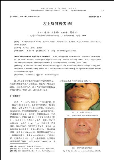
小涎腺涎石病较为罕见,且很多没有自觉症状,仅有极少数病例能够在初期就能正确诊断。涎石常使唾液排出受阻,并继发感染,造成腺体急性或反复发作的炎症。慢性期时应主要与黏液囊肿、副涎腺恶性或良性肿瘤、刺激性纤维瘤和黏膜下异物等鉴别诊断,急性期时应与感染性黏液囊肿、唇部蜂窝组织炎等鉴别诊断[2]。涎石的组织病理学表现为层板状低矿化的结节,位于唾液腺导管的远端;导管增生扩张,排泄管细胞出现鳞状化生;下方的组织内有淋巴细胞浸润;腺泡或多或少出现萎缩、消失[8]。
[参考文献]
[1]McGurk M, Escudier MP, Brown JE. Modern management of salivary calculi[J]. Br J Surg, 2005, 92(1):107-112.
[2]Ben Lagha N, Alantar A, Samson J, et al. Lithiasis of minor salivary glands: current data[J]. Oral Surg Oral Med Oral Pathol Oral Radiol Endod, 2005, 100(3):345-348.
[3]Alcure ML, Della Coletta R, Graner E, et al. Sialolithiasis of minor salivary glands: a clinical and histopathological study[J]. Gen Dent, 2005, 53(4):278-281.
[4]Grases F, Santiago C, Simonet BM, et al. Sialolithiasis: me-chanism of calculi formation and etiologic factors[J]. Clin Chim Acta, 2003, 334(1/2):131-136.
[5]Teymoortash A, Wollstein AC, Lippert BM, et al. Bacteria and pathogenesis of human salivary calculus[J]. Acta Oto-laryngol, 2002, 122(2):210-214.
[6]Huoh KC, Eisele DW. Etiologic factors in sialolithiasis[J]. Otolaryngol Head Neck Surg, 2011, 145(6):935-939.
[7]Alves de Matos AP, Carvalho PA, Almeida A, et al. On the structural diversity of sialoliths[J]. Microsc Microanal, 2007, 13(5):390-396.
[8]Lee LT, Wong YK. Pathogenesis and diverse histologic findings of sialolithiasis in minor salivary glands[J]. J Oral Maxillofac Surg, 2010, 68(2):465-470.
(本文编辑 李彩)
您现在查看是摘要介绍页,详见PDF附件。