空洞型肺结核的CT影像学特征及其与病理基础的关系
 |
| 第1页 |
参见附件。
doi:10.3969/j.issn.1007-614x.2014.14.54
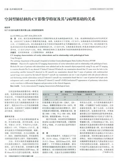
摘 要 目的:探讨空洞型肺结核的CT影像学特征及其与病理基础的关系。方法:收治肺结核患者36例作为研究对象,分别采用了X线和CT影像学进行检查。结果:X线显示17个空洞,CT 39个;X线检查显示空洞周围卫星病灶13例,CT观察到24例;经X线检查显示由于空洞而导致的肺内支气管播散患者3例,CT显示7例;经X线检查可见患者结核空洞并存在侧胸膜粘连以及增厚患者1例,CT显示3例;X线检查没有发现1例患者出现淋巴结肿大以及少量积液,CT显示2例(P<0.05)。结论:肺结核空洞的CT表现有助于临床结核病的鉴别诊断及防治。
关键词 空洞型肺结核 CT影像学特征 病理基础
CT imaging characteristics of cavity tuberculosis and its relationship with pathological basis
Yang Shengjun
The radiology department of the people's hospital in Leishan County,Qiandongnan State,Guizhou Province,557100
Abstract Objective:To explore the CT imaging characteristics of cavity tuberculosis and its relationship with pathological basis.Methods:36 cases of patients with tuberculosis were admited and as the research object,respectively using X-ray and CT imaging to examine.Results:The X-ray showed 17 holes,CT showed 39 holes;X-ray examination showed that 13 cases were the hole in the surrounding satellite lesions ......
您现在查看是摘要介绍页,详见PDF附件。