肝内胆管细胞癌的CT与MRI诊断(1)
 |
| 第1页 |
参见附件。
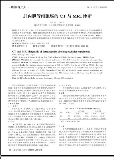
[摘要] 目的 探讨CT与MRI检查在肝内胆管细胞癌诊断中的临床应用价值。 方法 回顾性分析30例肝内胆管细胞癌患者的影像学资料。 结果 MRI定性诊断准确率为96.67%,CT定性诊断准确率为73.33%,两组定性诊断准确率比较,差异有统计学意义(P < 0.05),MRI与CT定位诊断准确率比较,差异无统计学意义(P > 0.05)。 结论 CT扫描与MRI检查都是肝内胆管细胞癌诊断与鉴别诊断的重要影像学手段,MRI检查更有利于病灶的早期发现与显示,值得临床推广。
[关键词] 胆管细胞癌;肝内;CT扫描;MRI检查
[中图分类号] R445 [文献标识码] A [文章编号] 1674―4721(2012)09(c)―0096―02
CT and MRI diagnosis of intrahepatic cholangiocellular carcinoma
GONG Wensong LIU Yongjiu
CT/MRI Department of Jingmen Municipal First People''s Hospital in Hubei Province, Jingmen 448000, China
[Abstract] Objective To investigate the clinical application of CT, MRI testing for intrahepatic cholangiocellular carcinoma. Methods The imaging data of 30 cases with intrahepatic cholangiocellular carcinoma were retrospectively analyzed. Results The qualitative diagnosis accuracy rate of MRI was 96.67%, while the rate of CT was 73.33%, there was significant difference between two groups (P < 0.05). And level diagnosis rate of CT and MRI, there was no significant difference between two groups (P > 0.05). Conclusion CT and MRI are important imaging methods to diagnosis and antidiastole the intrahepatic cholangiocellular carcinoma, while MRI testing is better to detect and reveal the focus in early stage, is worthy for being widely used in treatment.
[Key words] Cholangiocellular carcinoma; Intrahepatic; CT scan; MRI examination
肝内胆管细胞癌(PPC)是指起源于二级胆管在内的末梢肝内小胆管的胆管内皮细胞的原发性恶性肿瘤[1],发病率占所有胆管细胞癌的5%~10%[2],其临床表现复杂多样,术前误诊率较高。为探讨CT扫描与MRI检查在肝内胆管细胞癌诊断中的临床应用价值,笔者对2009年1月~2011年12月在本院经临床手术病理确诊的30例肝内胆管细胞癌患者的影像学资料进行了回顾性分析,现将研究结果总结报道如下:
1 资料与方法
1.1 一般资料
本组共30例,男17例,女13例,年龄32~73岁,平均52岁,其中因上腹隐痛不适、乏力消瘦就诊发现6例,上腹肿块就诊发现6例,因慢性黄疸就诊发现15例,正常体检发现3例,实验室检查甲胎蛋白(AFP)均为阴性,癌胚抗原(CEA)及CA125阳性17例 ......
您现在查看是摘要介绍页,详见PDF附件(2068kb)。